Chlorine »
PDB 3lz1-3m98 »
3m1y »
Chlorine in PDB 3m1y: Crystal Structure of A Phosphoserine Phosphatase (Serb) From Helicobacter Pylori
Protein crystallography data
The structure of Crystal Structure of A Phosphoserine Phosphatase (Serb) From Helicobacter Pylori, PDB code: 3m1y
was solved by
B.Syed Ibrahim,
S.K.Burley,
S.Swaminathan,
New York Sgxresearch Center For Structural Genomics (Nysgxrc),
with X-Ray Crystallography technique. A brief refinement statistics is given in the table below:
| Resolution Low / High (Å) | 44.07 / 2.40 |
| Space group | P 21 21 21 |
| Cell size a, b, c (Å), α, β, γ (°) | 51.607, 84.719, 204.826, 90.00, 90.00, 90.00 |
| R / Rfree (%) | 22.9 / 27.1 |
Other elements in 3m1y:
The structure of Crystal Structure of A Phosphoserine Phosphatase (Serb) From Helicobacter Pylori also contains other interesting chemical elements:
| Magnesium | (Mg) | 5 atoms |
Chlorine Binding Sites:
The binding sites of Chlorine atom in the Crystal Structure of A Phosphoserine Phosphatase (Serb) From Helicobacter Pylori
(pdb code 3m1y). This binding sites where shown within
5.0 Angstroms radius around Chlorine atom.
In total 5 binding sites of Chlorine where determined in the Crystal Structure of A Phosphoserine Phosphatase (Serb) From Helicobacter Pylori, PDB code: 3m1y:
Jump to Chlorine binding site number: 1; 2; 3; 4; 5;
In total 5 binding sites of Chlorine where determined in the Crystal Structure of A Phosphoserine Phosphatase (Serb) From Helicobacter Pylori, PDB code: 3m1y:
Jump to Chlorine binding site number: 1; 2; 3; 4; 5;
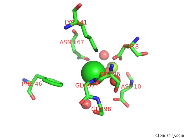
Chlorine binding site 1 out of 5 in 3m1y
Go back to
Chlorine binding site 1 out
of 5 in the Crystal Structure of A Phosphoserine Phosphatase (Serb) From Helicobacter Pylori
Mono view
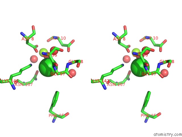
Stereo pair view
Mono view
Stereo pair view
A full contact list of Chlorine with other atoms in the Cl binding
site number 1 of Crystal Structure of A Phosphoserine Phosphatase (Serb) From Helicobacter Pylori within 5.0Å range:
|
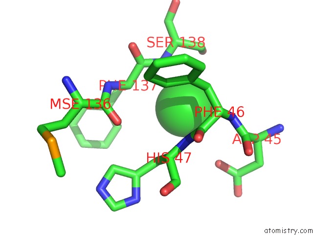
Chlorine binding site 2 out of 5 in 3m1y
Go back to
Chlorine binding site 2 out
of 5 in the Crystal Structure of A Phosphoserine Phosphatase (Serb) From Helicobacter Pylori
Mono view

Stereo pair view
Mono view
Stereo pair view
A full contact list of Chlorine with other atoms in the Cl binding
site number 2 of Crystal Structure of A Phosphoserine Phosphatase (Serb) From Helicobacter Pylori within 5.0Å range:
|
Chlorine binding site 3 out of 5 in 3m1y
Go back to
Chlorine binding site 3 out
of 5 in the Crystal Structure of A Phosphoserine Phosphatase (Serb) From Helicobacter Pylori

Mono view

Stereo pair view
Mono view
Stereo pair view
A full contact list of Chlorine with other atoms in the Cl binding
site number 3 of Crystal Structure of A Phosphoserine Phosphatase (Serb) From Helicobacter Pylori within 5.0Å range:
|
Chlorine binding site 4 out of 5 in 3m1y
Go back to
Chlorine binding site 4 out
of 5 in the Crystal Structure of A Phosphoserine Phosphatase (Serb) From Helicobacter Pylori

Mono view

Stereo pair view
Mono view
Stereo pair view
A full contact list of Chlorine with other atoms in the Cl binding
site number 4 of Crystal Structure of A Phosphoserine Phosphatase (Serb) From Helicobacter Pylori within 5.0Å range:
|
Chlorine binding site 5 out of 5 in 3m1y
Go back to
Chlorine binding site 5 out
of 5 in the Crystal Structure of A Phosphoserine Phosphatase (Serb) From Helicobacter Pylori

Mono view

Stereo pair view
Mono view
Stereo pair view
A full contact list of Chlorine with other atoms in the Cl binding
site number 5 of Crystal Structure of A Phosphoserine Phosphatase (Serb) From Helicobacter Pylori within 5.0Å range:
|
Reference:
B.Syed Ibrahim,
S.K.Burley,
S.Swaminathan.
Crystal Structure of A Phosphoserine Phosphatase (Serb) From Helicobacter Pylori To Be Published.
Page generated: Fri Jul 11 07:44:08 2025
Last articles
I in 4S22I in 4S2H
I in 4QX5
I in 4S2G
I in 4S2F
I in 4RX1
I in 4S2D
I in 4RYM
I in 4REC
I in 4RCF