Chlorine »
PDB 3lz1-3m98 »
3m6z »
Chlorine in PDB 3m6z: Crystal Structure of An N-Terminal 44 kDa Fragment of Topoisomerase V in the Presence of Guanidium Hydrochloride
Protein crystallography data
The structure of Crystal Structure of An N-Terminal 44 kDa Fragment of Topoisomerase V in the Presence of Guanidium Hydrochloride, PDB code: 3m6z
was solved by
R.Rajan,
B.Taneja,
A.Mondragon,
with X-Ray Crystallography technique. A brief refinement statistics is given in the table below:
| Resolution Low / High (Å) | 28.90 / 1.40 |
| Space group | P 21 21 21 |
| Cell size a, b, c (Å), α, β, γ (°) | 63.640, 80.110, 137.220, 90.00, 90.00, 90.00 |
| R / Rfree (%) | 16.5 / 18.4 |
Other elements in 3m6z:
The structure of Crystal Structure of An N-Terminal 44 kDa Fragment of Topoisomerase V in the Presence of Guanidium Hydrochloride also contains other interesting chemical elements:
| Magnesium | (Mg) | 3 atoms |
Chlorine Binding Sites:
The binding sites of Chlorine atom in the Crystal Structure of An N-Terminal 44 kDa Fragment of Topoisomerase V in the Presence of Guanidium Hydrochloride
(pdb code 3m6z). This binding sites where shown within
5.0 Angstroms radius around Chlorine atom.
In total 2 binding sites of Chlorine where determined in the Crystal Structure of An N-Terminal 44 kDa Fragment of Topoisomerase V in the Presence of Guanidium Hydrochloride, PDB code: 3m6z:
Jump to Chlorine binding site number: 1; 2;
In total 2 binding sites of Chlorine where determined in the Crystal Structure of An N-Terminal 44 kDa Fragment of Topoisomerase V in the Presence of Guanidium Hydrochloride, PDB code: 3m6z:
Jump to Chlorine binding site number: 1; 2;
Chlorine binding site 1 out of 2 in 3m6z
Go back to
Chlorine binding site 1 out
of 2 in the Crystal Structure of An N-Terminal 44 kDa Fragment of Topoisomerase V in the Presence of Guanidium Hydrochloride
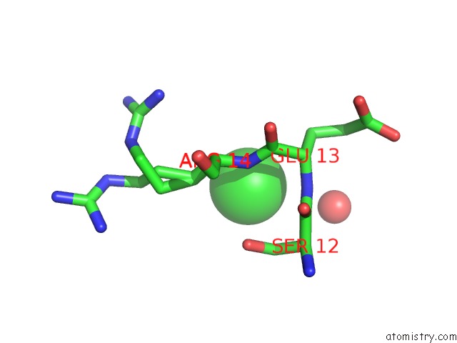
Mono view
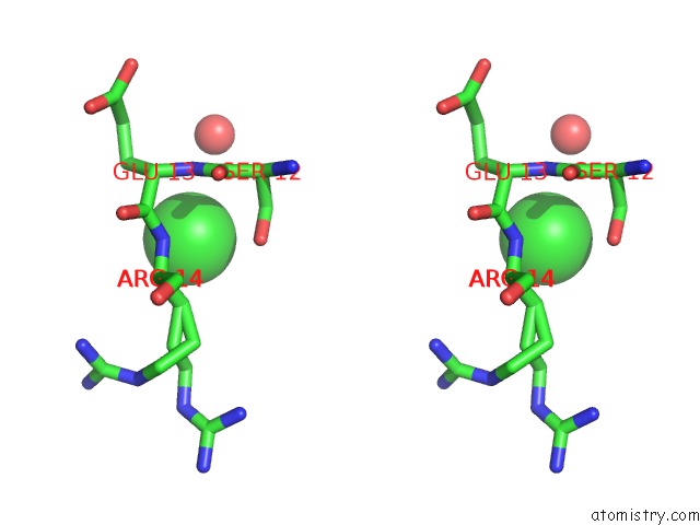
Stereo pair view
Mono view
Stereo pair view
A full contact list of Chlorine with other atoms in the Cl binding
site number 1 of Crystal Structure of An N-Terminal 44 kDa Fragment of Topoisomerase V in the Presence of Guanidium Hydrochloride within 5.0Å range:
|
Chlorine binding site 2 out of 2 in 3m6z
Go back to
Chlorine binding site 2 out
of 2 in the Crystal Structure of An N-Terminal 44 kDa Fragment of Topoisomerase V in the Presence of Guanidium Hydrochloride
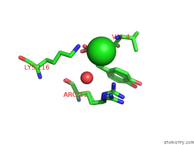
Mono view
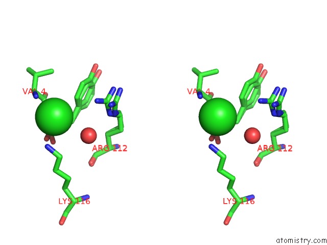
Stereo pair view
Mono view
Stereo pair view
A full contact list of Chlorine with other atoms in the Cl binding
site number 2 of Crystal Structure of An N-Terminal 44 kDa Fragment of Topoisomerase V in the Presence of Guanidium Hydrochloride within 5.0Å range:
|
Reference:
R.Rajan,
B.Taneja,
A.Mondragon.
Structures of Minimal Catalytic Fragments of Topoisomerase V Reveals Conformational Changes Relevant For Dna Binding. Structure V. 18 829 2010.
ISSN: ISSN 0969-2126
PubMed: 20637419
DOI: 10.1016/J.STR.2010.03.006
Page generated: Fri Jul 11 07:47:01 2025
ISSN: ISSN 0969-2126
PubMed: 20637419
DOI: 10.1016/J.STR.2010.03.006
Last articles
I in 4P9TI in 4PNX
I in 4PVQ
I in 4PVG
I in 4PGC
I in 4PNS
I in 4P4Z
I in 4P1E
I in 4P4X
I in 4P4Y