Chlorine »
PDB 3mvu-3n4a »
3n3t »
Chlorine in PDB 3n3t: Crystal Structure of Putative Diguanylate Cyclase/Phosphodiesterase Complex with Cyclic Di-Gmp
Protein crystallography data
The structure of Crystal Structure of Putative Diguanylate Cyclase/Phosphodiesterase Complex with Cyclic Di-Gmp, PDB code: 3n3t
was solved by
C.Chang,
X.Xu,
H.Cui,
A.Savchenko,
A.Edwards,
A.Joachimiak,
Midwest Centerfor Structural Genomics (Mcsg),
with X-Ray Crystallography technique. A brief refinement statistics is given in the table below:
| Resolution Low / High (Å) | 39.96 / 2.35 |
| Space group | P 21 21 21 |
| Cell size a, b, c (Å), α, β, γ (°) | 51.560, 63.201, 173.987, 90.00, 90.00, 90.00 |
| R / Rfree (%) | 18.4 / 25.4 |
Other elements in 3n3t:
The structure of Crystal Structure of Putative Diguanylate Cyclase/Phosphodiesterase Complex with Cyclic Di-Gmp also contains other interesting chemical elements:
| Magnesium | (Mg) | 4 atoms |
Chlorine Binding Sites:
The binding sites of Chlorine atom in the Crystal Structure of Putative Diguanylate Cyclase/Phosphodiesterase Complex with Cyclic Di-Gmp
(pdb code 3n3t). This binding sites where shown within
5.0 Angstroms radius around Chlorine atom.
In total only one binding site of Chlorine was determined in the Crystal Structure of Putative Diguanylate Cyclase/Phosphodiesterase Complex with Cyclic Di-Gmp, PDB code: 3n3t:
In total only one binding site of Chlorine was determined in the Crystal Structure of Putative Diguanylate Cyclase/Phosphodiesterase Complex with Cyclic Di-Gmp, PDB code: 3n3t:
Chlorine binding site 1 out of 1 in 3n3t
Go back to
Chlorine binding site 1 out
of 1 in the Crystal Structure of Putative Diguanylate Cyclase/Phosphodiesterase Complex with Cyclic Di-Gmp
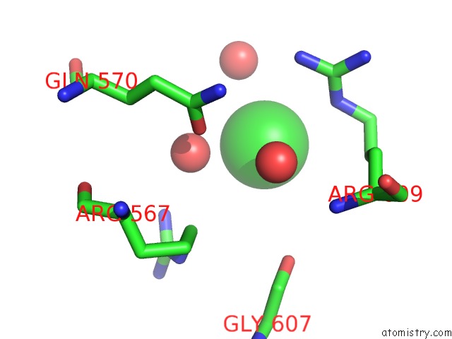
Mono view
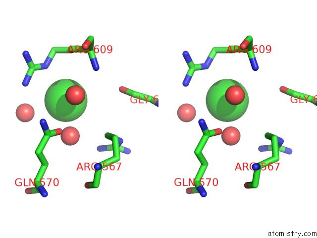
Stereo pair view
Mono view
Stereo pair view
A full contact list of Chlorine with other atoms in the Cl binding
site number 1 of Crystal Structure of Putative Diguanylate Cyclase/Phosphodiesterase Complex with Cyclic Di-Gmp within 5.0Å range:
|
Reference:
A.Tchigvintsev,
X.Xu,
A.Singer,
C.Chang,
G.Brown,
M.Proudfoot,
H.Cui,
R.Flick,
W.F.Anderson,
A.Joachimiak,
M.Y.Galperin,
A.Savchenko,
A.F.Yakunin.
Structural Insight Into the Mechanism of C-Di-Gmp Hydrolysis By Eal Domain Phosphodiesterases. J.Mol.Biol. V. 402 524 2010.
ISSN: ISSN 0022-2836
PubMed: 20691189
DOI: 10.1016/J.JMB.2010.07.050
Page generated: Fri Jul 11 08:04:32 2025
ISSN: ISSN 0022-2836
PubMed: 20691189
DOI: 10.1016/J.JMB.2010.07.050
Last articles
Mg in 5OEAMg in 5OET
Mg in 5OED
Mg in 5OEB
Mg in 5OEC
Mg in 5ODZ
Mg in 5ODO
Mg in 5ODC
Mg in 5ODJ
Mg in 5OCT