Chlorine »
PDB 3nbu-3nnw »
3nd0 »
Chlorine in PDB 3nd0: X-Ray Crystal Structure of A Slow Cyanobacterial Cl-/H+ Antiporter
Protein crystallography data
The structure of X-Ray Crystal Structure of A Slow Cyanobacterial Cl-/H+ Antiporter, PDB code: 3nd0
was solved by
H.Jayaram,
J.Robertson,
F.Wu,
C.Williams,
C.Miller,
with X-Ray Crystallography technique. A brief refinement statistics is given in the table below:
| Resolution Low / High (Å) | 65.22 / 3.20 |
| Space group | P 3 2 1 |
| Cell size a, b, c (Å), α, β, γ (°) | 203.790, 203.790, 96.820, 90.00, 90.00, 120.00 |
| R / Rfree (%) | 24.3 / 26.9 |
Chlorine Binding Sites:
The binding sites of Chlorine atom in the X-Ray Crystal Structure of A Slow Cyanobacterial Cl-/H+ Antiporter
(pdb code 3nd0). This binding sites where shown within
5.0 Angstroms radius around Chlorine atom.
In total 2 binding sites of Chlorine where determined in the X-Ray Crystal Structure of A Slow Cyanobacterial Cl-/H+ Antiporter, PDB code: 3nd0:
Jump to Chlorine binding site number: 1; 2;
In total 2 binding sites of Chlorine where determined in the X-Ray Crystal Structure of A Slow Cyanobacterial Cl-/H+ Antiporter, PDB code: 3nd0:
Jump to Chlorine binding site number: 1; 2;
Chlorine binding site 1 out of 2 in 3nd0
Go back to
Chlorine binding site 1 out
of 2 in the X-Ray Crystal Structure of A Slow Cyanobacterial Cl-/H+ Antiporter
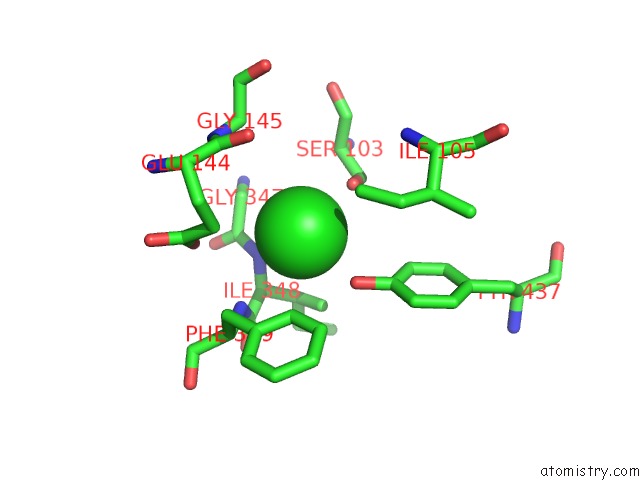
Mono view
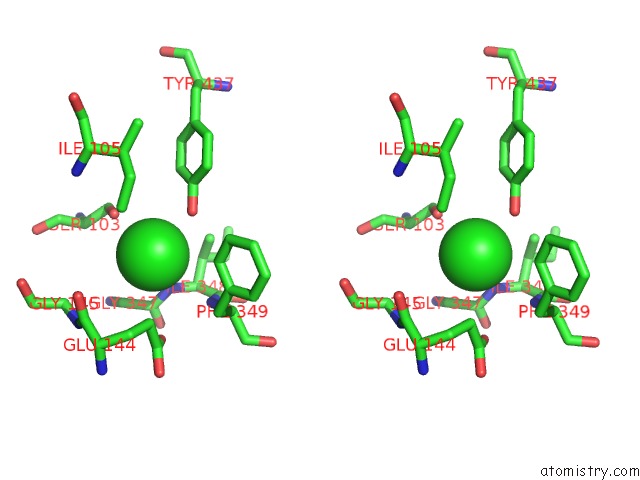
Stereo pair view
Mono view
Stereo pair view
A full contact list of Chlorine with other atoms in the Cl binding
site number 1 of X-Ray Crystal Structure of A Slow Cyanobacterial Cl-/H+ Antiporter within 5.0Å range:
|
Chlorine binding site 2 out of 2 in 3nd0
Go back to
Chlorine binding site 2 out
of 2 in the X-Ray Crystal Structure of A Slow Cyanobacterial Cl-/H+ Antiporter
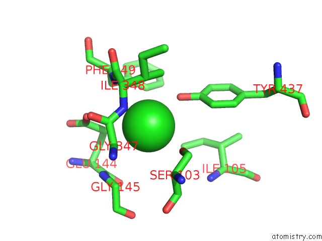
Mono view
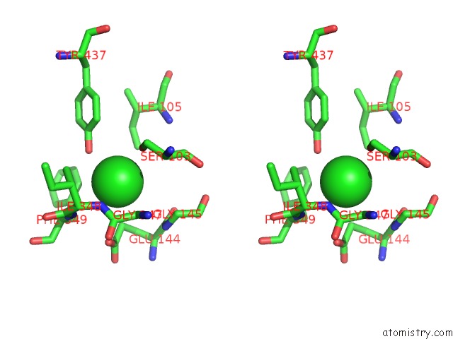
Stereo pair view
Mono view
Stereo pair view
A full contact list of Chlorine with other atoms in the Cl binding
site number 2 of X-Ray Crystal Structure of A Slow Cyanobacterial Cl-/H+ Antiporter within 5.0Å range:
|
Reference:
H.Jayaram,
J.L.Robertson,
F.Wu,
C.Williams,
C.Miller.
Structure of A Slow Clc Cl(-)/H(+) Antiporter From A Cyanobacterium. Biochemistry V. 50 788 2011.
ISSN: ISSN 0006-2960
PubMed: 21174448
DOI: 10.1021/BI1019258
Page generated: Fri Jul 11 08:18:56 2025
ISSN: ISSN 0006-2960
PubMed: 21174448
DOI: 10.1021/BI1019258
Last articles
Mn in 3WU2Mn in 3WQO
Mn in 3WKJ
Mn in 3WOG
Mn in 3WRO
Mn in 3WCS
Mn in 3WEI
Mn in 3WI8
Mn in 3WAK
Mn in 3W99