Chlorine »
PDB 3nbu-3nnw »
3nks »
Chlorine in PDB 3nks: Structure of Human Protoporphyrinogen IX Oxidase
Enzymatic activity of Structure of Human Protoporphyrinogen IX Oxidase
All present enzymatic activity of Structure of Human Protoporphyrinogen IX Oxidase:
1.3.3.4;
1.3.3.4;
Protein crystallography data
The structure of Structure of Human Protoporphyrinogen IX Oxidase, PDB code: 3nks
was solved by
Y.Shen,
with X-Ray Crystallography technique. A brief refinement statistics is given in the table below:
| Resolution Low / High (Å) | 29.71 / 1.90 |
| Space group | H 3 2 |
| Cell size a, b, c (Å), α, β, γ (°) | 136.702, 136.702, 158.877, 90.00, 90.00, 120.00 |
| R / Rfree (%) | 20.6 / 23.2 |
Other elements in 3nks:
The structure of Structure of Human Protoporphyrinogen IX Oxidase also contains other interesting chemical elements:
| Fluorine | (F) | 3 atoms |
Chlorine Binding Sites:
The binding sites of Chlorine atom in the Structure of Human Protoporphyrinogen IX Oxidase
(pdb code 3nks). This binding sites where shown within
5.0 Angstroms radius around Chlorine atom.
In total only one binding site of Chlorine was determined in the Structure of Human Protoporphyrinogen IX Oxidase, PDB code: 3nks:
In total only one binding site of Chlorine was determined in the Structure of Human Protoporphyrinogen IX Oxidase, PDB code: 3nks:
Chlorine binding site 1 out of 1 in 3nks
Go back to
Chlorine binding site 1 out
of 1 in the Structure of Human Protoporphyrinogen IX Oxidase
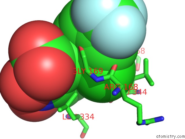
Mono view
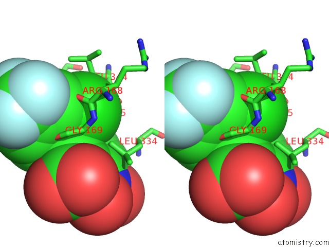
Stereo pair view
Mono view
Stereo pair view
A full contact list of Chlorine with other atoms in the Cl binding
site number 1 of Structure of Human Protoporphyrinogen IX Oxidase within 5.0Å range:
|
Reference:
X.Qin,
Y.Tan,
L.Wang,
Z.Wang,
B.Wang,
X.Wen,
G.Yang,
Z.Xi,
Y.Shen.
Structural Insight Into Human Variegate Porphyria Disease Faseb J. V. 25 653 2011.
ISSN: ISSN 0892-6638
PubMed: 21048046
DOI: 10.1096/FJ.10-170811
Page generated: Fri Jul 11 08:22:29 2025
ISSN: ISSN 0892-6638
PubMed: 21048046
DOI: 10.1096/FJ.10-170811
Last articles
Mn in 4QS5Mn in 4QRN
Mn in 4QPX
Mn in 4QKD
Mn in 4QKB
Mn in 4QNJ
Mn in 4QKF
Mn in 4QKN
Mn in 4Q42
Mn in 4Q41