Chlorine »
PDB 3nnx-3nw8 »
3nuq »
Chlorine in PDB 3nuq: Structure of A Putative Nucleotide Phosphatase From Saccharomyces Cerevisiae
Enzymatic activity of Structure of A Putative Nucleotide Phosphatase From Saccharomyces Cerevisiae
All present enzymatic activity of Structure of A Putative Nucleotide Phosphatase From Saccharomyces Cerevisiae:
3.1.3.5;
3.1.3.5;
Protein crystallography data
The structure of Structure of A Putative Nucleotide Phosphatase From Saccharomyces Cerevisiae, PDB code: 3nuq
was solved by
A.Dong,
C.Yang,
A.U.Singer,
E.Evdokimova,
M.Kudritsdka,
G.Brown,
A.M.Edwards,
A.Joachimiak,
A.Savchenko,
A.F.Yakunin,
Midwest Center Forstructural Genomics (Mcsg),
with X-Ray Crystallography technique. A brief refinement statistics is given in the table below:
| Resolution Low / High (Å) | 23.38 / 1.70 |
| Space group | P 21 21 21 |
| Cell size a, b, c (Å), α, β, γ (°) | 58.474, 64.664, 67.708, 90.00, 90.00, 90.00 |
| R / Rfree (%) | 19.4 / 22.6 |
Other elements in 3nuq:
The structure of Structure of A Putative Nucleotide Phosphatase From Saccharomyces Cerevisiae also contains other interesting chemical elements:
| Sodium | (Na) | 1 atom |
Chlorine Binding Sites:
The binding sites of Chlorine atom in the Structure of A Putative Nucleotide Phosphatase From Saccharomyces Cerevisiae
(pdb code 3nuq). This binding sites where shown within
5.0 Angstroms radius around Chlorine atom.
In total 2 binding sites of Chlorine where determined in the Structure of A Putative Nucleotide Phosphatase From Saccharomyces Cerevisiae, PDB code: 3nuq:
Jump to Chlorine binding site number: 1; 2;
In total 2 binding sites of Chlorine where determined in the Structure of A Putative Nucleotide Phosphatase From Saccharomyces Cerevisiae, PDB code: 3nuq:
Jump to Chlorine binding site number: 1; 2;
Chlorine binding site 1 out of 2 in 3nuq
Go back to
Chlorine binding site 1 out
of 2 in the Structure of A Putative Nucleotide Phosphatase From Saccharomyces Cerevisiae
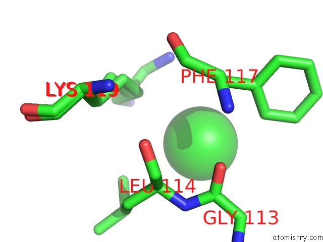
Mono view
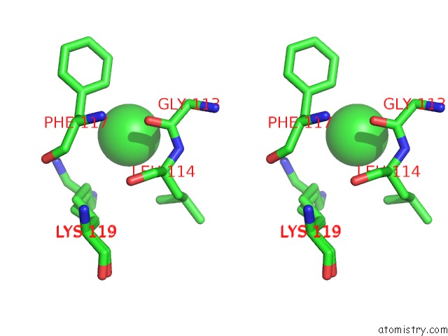
Stereo pair view
Mono view
Stereo pair view
A full contact list of Chlorine with other atoms in the Cl binding
site number 1 of Structure of A Putative Nucleotide Phosphatase From Saccharomyces Cerevisiae within 5.0Å range:
|
Chlorine binding site 2 out of 2 in 3nuq
Go back to
Chlorine binding site 2 out
of 2 in the Structure of A Putative Nucleotide Phosphatase From Saccharomyces Cerevisiae
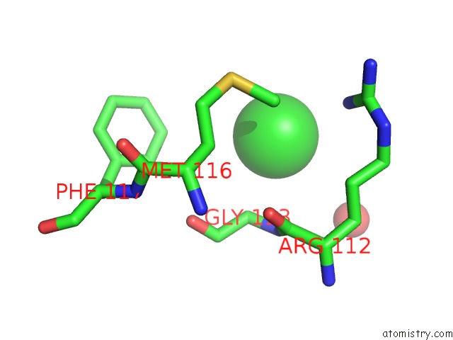
Mono view
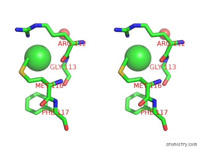
Stereo pair view
Mono view
Stereo pair view
A full contact list of Chlorine with other atoms in the Cl binding
site number 2 of Structure of A Putative Nucleotide Phosphatase From Saccharomyces Cerevisiae within 5.0Å range:
|
Reference:
G.Brown,
E.Evdokimova,
M.Kudritska,
A.Dong,
C.Yang,
A.U.Singer,
A.M.Edwards,
A.Savchenko,
A.F.Yakunin.
Structure of A Putative Nucleotide Phosphatase From Saccharomyces Cerevisiae To Be Published.
Page generated: Fri Jul 11 08:29:31 2025
Last articles
Fe in 4UN1Fe in 4ULV
Fe in 4UMZ
Fe in 4UHL
Fe in 4UHX
Fe in 4UHW
Fe in 4UII
Fe in 4UHI
Fe in 4UHA
Fe in 4UH9