Chlorine »
PDB 3nw9-3o6j »
3o32 »
Chlorine in PDB 3o32: Crystal Structure of 4-Chlorocatechol Dioxygenase From Rhodococcus Opacus 1CP in Complex with 3,5-Dichlorocatechol
Protein crystallography data
The structure of Crystal Structure of 4-Chlorocatechol Dioxygenase From Rhodococcus Opacus 1CP in Complex with 3,5-Dichlorocatechol, PDB code: 3o32
was solved by
M.Ferraroni,
F.Briganti,
M.Kolomytseva,
L.Golovleva,
with X-Ray Crystallography technique. A brief refinement statistics is given in the table below:
| Resolution Low / High (Å) | 15.00 / 2.85 |
| Space group | P 63 2 2 |
| Cell size a, b, c (Å), α, β, γ (°) | 90.530, 90.530, 309.830, 90.00, 90.00, 120.00 |
| R / Rfree (%) | 21.3 / 28.9 |
Other elements in 3o32:
The structure of Crystal Structure of 4-Chlorocatechol Dioxygenase From Rhodococcus Opacus 1CP in Complex with 3,5-Dichlorocatechol also contains other interesting chemical elements:
| Iron | (Fe) | 2 atoms |
Chlorine Binding Sites:
The binding sites of Chlorine atom in the Crystal Structure of 4-Chlorocatechol Dioxygenase From Rhodococcus Opacus 1CP in Complex with 3,5-Dichlorocatechol
(pdb code 3o32). This binding sites where shown within
5.0 Angstroms radius around Chlorine atom.
In total 2 binding sites of Chlorine where determined in the Crystal Structure of 4-Chlorocatechol Dioxygenase From Rhodococcus Opacus 1CP in Complex with 3,5-Dichlorocatechol, PDB code: 3o32:
Jump to Chlorine binding site number: 1; 2;
In total 2 binding sites of Chlorine where determined in the Crystal Structure of 4-Chlorocatechol Dioxygenase From Rhodococcus Opacus 1CP in Complex with 3,5-Dichlorocatechol, PDB code: 3o32:
Jump to Chlorine binding site number: 1; 2;
Chlorine binding site 1 out of 2 in 3o32
Go back to
Chlorine binding site 1 out
of 2 in the Crystal Structure of 4-Chlorocatechol Dioxygenase From Rhodococcus Opacus 1CP in Complex with 3,5-Dichlorocatechol
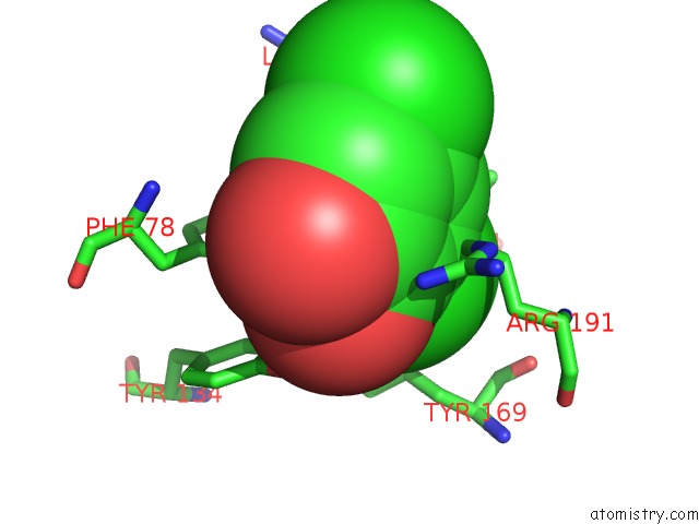
Mono view
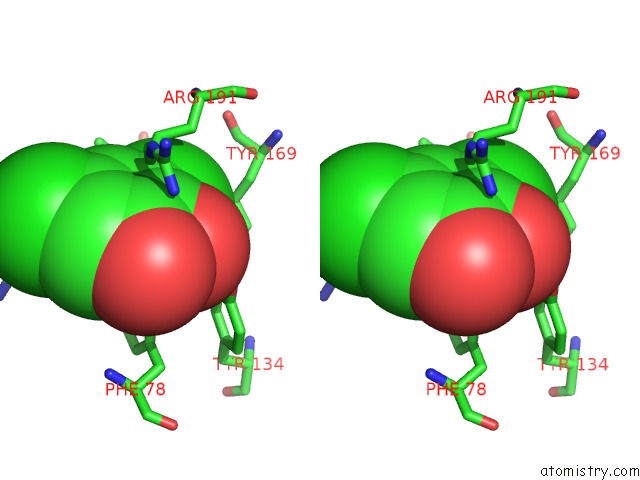
Stereo pair view
Mono view
Stereo pair view
A full contact list of Chlorine with other atoms in the Cl binding
site number 1 of Crystal Structure of 4-Chlorocatechol Dioxygenase From Rhodococcus Opacus 1CP in Complex with 3,5-Dichlorocatechol within 5.0Å range:
|
Chlorine binding site 2 out of 2 in 3o32
Go back to
Chlorine binding site 2 out
of 2 in the Crystal Structure of 4-Chlorocatechol Dioxygenase From Rhodococcus Opacus 1CP in Complex with 3,5-Dichlorocatechol
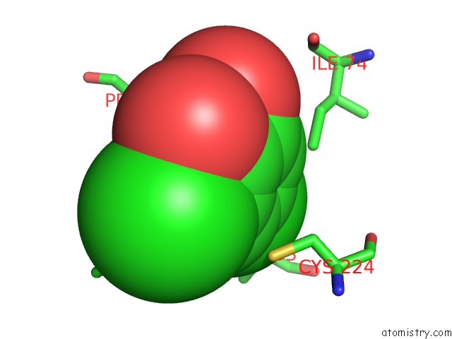
Mono view
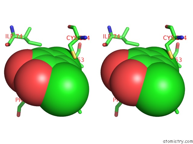
Stereo pair view
Mono view
Stereo pair view
A full contact list of Chlorine with other atoms in the Cl binding
site number 2 of Crystal Structure of 4-Chlorocatechol Dioxygenase From Rhodococcus Opacus 1CP in Complex with 3,5-Dichlorocatechol within 5.0Å range:
|
Reference:
M.Ferraroni,
M.Kolomytseva,
A.Scozzafava,
L.Golovleva,
F.Briganti.
X-Ray Structures of 4-Chlorocatechol 1,2-Dioxygenase Adducts with Substituted Catechols: New Perspectives in the Molecular Basis of Intradiol Ring Cleaving Dioxygenases Specificity. J. Struct. Biol. V. 181 274 2013.
ISSN: ESSN 1095-8657
PubMed: 23261399
DOI: 10.1016/J.JSB.2012.11.007
Page generated: Fri Jul 11 08:32:29 2025
ISSN: ESSN 1095-8657
PubMed: 23261399
DOI: 10.1016/J.JSB.2012.11.007
Last articles
Na in 1VI6Na in 1VKG
Na in 1VMJ
Na in 1VMH
Na in 1VMF
Na in 1VLM
Na in 1VK1
Na in 1VIZ
Na in 1VEL
Na in 1VE8