Chlorine »
PDB 3pf6-3pne »
3pm5 »
Chlorine in PDB 3pm5: Crystal Structure of Boxb in Mixed Valent State with Bound Benzoyl-Coa
Enzymatic activity of Crystal Structure of Boxb in Mixed Valent State with Bound Benzoyl-Coa
All present enzymatic activity of Crystal Structure of Boxb in Mixed Valent State with Bound Benzoyl-Coa:
1.14.12.21;
1.14.12.21;
Protein crystallography data
The structure of Crystal Structure of Boxb in Mixed Valent State with Bound Benzoyl-Coa, PDB code: 3pm5
was solved by
T.Weinert,
L.Rather,
G.Fuchs,
U.Ermler,
with X-Ray Crystallography technique. A brief refinement statistics is given in the table below:
| Resolution Low / High (Å) | 49.40 / 2.30 |
| Space group | C 1 2 1 |
| Cell size a, b, c (Å), α, β, γ (°) | 208.180, 76.650, 148.200, 90.00, 108.34, 90.00 |
| R / Rfree (%) | 17.4 / 22.4 |
Other elements in 3pm5:
The structure of Crystal Structure of Boxb in Mixed Valent State with Bound Benzoyl-Coa also contains other interesting chemical elements:
| Iron | (Fe) | 8 atoms |
Chlorine Binding Sites:
The binding sites of Chlorine atom in the Crystal Structure of Boxb in Mixed Valent State with Bound Benzoyl-Coa
(pdb code 3pm5). This binding sites where shown within
5.0 Angstroms radius around Chlorine atom.
In total 4 binding sites of Chlorine where determined in the Crystal Structure of Boxb in Mixed Valent State with Bound Benzoyl-Coa, PDB code: 3pm5:
Jump to Chlorine binding site number: 1; 2; 3; 4;
In total 4 binding sites of Chlorine where determined in the Crystal Structure of Boxb in Mixed Valent State with Bound Benzoyl-Coa, PDB code: 3pm5:
Jump to Chlorine binding site number: 1; 2; 3; 4;
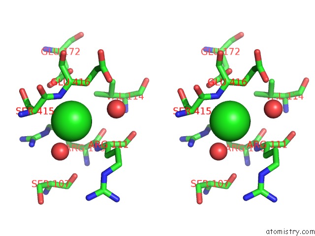
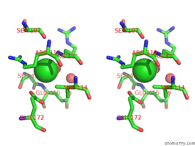
Chlorine binding site 1 out of 4 in 3pm5
Go back to
Chlorine binding site 1 out
of 4 in the Crystal Structure of Boxb in Mixed Valent State with Bound Benzoyl-Coa
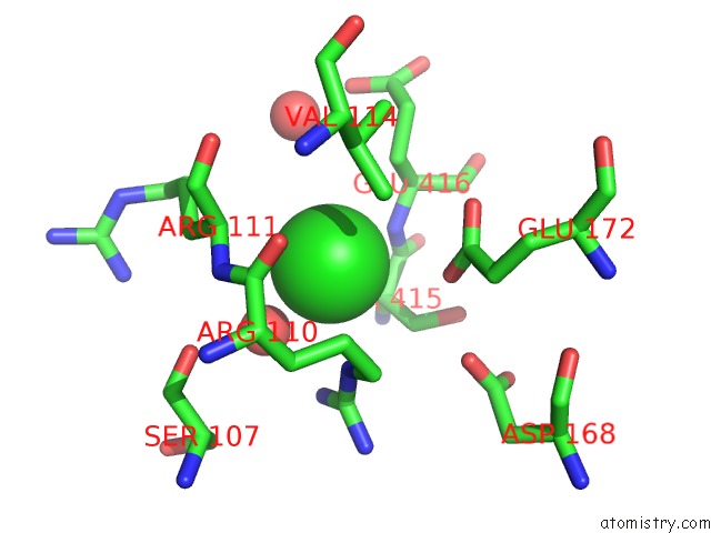
Mono view

Stereo pair view
Mono view
Stereo pair view
A full contact list of Chlorine with other atoms in the Cl binding
site number 1 of Crystal Structure of Boxb in Mixed Valent State with Bound Benzoyl-Coa within 5.0Å range:
|
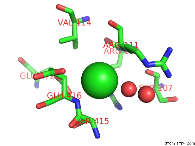
Chlorine binding site 2 out of 4 in 3pm5
Go back to
Chlorine binding site 2 out
of 4 in the Crystal Structure of Boxb in Mixed Valent State with Bound Benzoyl-Coa

Mono view
Stereo pair view
Mono view
Stereo pair view
A full contact list of Chlorine with other atoms in the Cl binding
site number 2 of Crystal Structure of Boxb in Mixed Valent State with Bound Benzoyl-Coa within 5.0Å range:
|
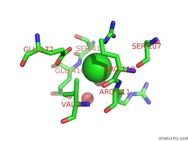
Chlorine binding site 3 out of 4 in 3pm5
Go back to
Chlorine binding site 3 out
of 4 in the Crystal Structure of Boxb in Mixed Valent State with Bound Benzoyl-Coa
Mono view

Stereo pair view
Mono view
Stereo pair view
A full contact list of Chlorine with other atoms in the Cl binding
site number 3 of Crystal Structure of Boxb in Mixed Valent State with Bound Benzoyl-Coa within 5.0Å range:
|
Chlorine binding site 4 out of 4 in 3pm5
Go back to
Chlorine binding site 4 out
of 4 in the Crystal Structure of Boxb in Mixed Valent State with Bound Benzoyl-Coa
Mono view
Stereo pair view
Mono view
Stereo pair view
A full contact list of Chlorine with other atoms in the Cl binding
site number 4 of Crystal Structure of Boxb in Mixed Valent State with Bound Benzoyl-Coa within 5.0Å range:
|
Reference:
L.J.Rather,
T.Weinert,
U.Demmer,
E.Bill,
W.Ismail,
G.Fuchs,
U.Ermler.
Structure and Mechanism of the Diiron Benzoyl-Coenzyme A Epoxidase Boxb. J.Biol.Chem. V. 286 29241 2011.
ISSN: ISSN 0021-9258
PubMed: 21632537
DOI: 10.1074/JBC.M111.236893
Page generated: Fri Jul 11 09:09:57 2025
ISSN: ISSN 0021-9258
PubMed: 21632537
DOI: 10.1074/JBC.M111.236893
Last articles
Fe in 2YXOFe in 2YRS
Fe in 2YXC
Fe in 2YNM
Fe in 2YVJ
Fe in 2YP1
Fe in 2YU2
Fe in 2YU1
Fe in 2YQB
Fe in 2YOO