Chlorine »
PDB 3pf6-3pne »
3pmo »
Chlorine in PDB 3pmo: The Structure of Lpxd From Pseudomonas Aeruginosa at 1.3 A Resolution
Protein crystallography data
The structure of The Structure of Lpxd From Pseudomonas Aeruginosa at 1.3 A Resolution, PDB code: 3pmo
was solved by
J.Badger,
B.Chie-Leon,
C.Logan,
V.Sridhar,
B.Sankaran,
P.H.Zwart,
V.Nienaber,
with X-Ray Crystallography technique. A brief refinement statistics is given in the table below:
| Resolution Low / High (Å) | 26.85 / 1.30 |
| Space group | H 3 |
| Cell size a, b, c (Å), α, β, γ (°) | 106.190, 106.190, 93.380, 90.00, 90.00, 120.00 |
| R / Rfree (%) | 16.4 / 18.5 |
Chlorine Binding Sites:
The binding sites of Chlorine atom in the The Structure of Lpxd From Pseudomonas Aeruginosa at 1.3 A Resolution
(pdb code 3pmo). This binding sites where shown within
5.0 Angstroms radius around Chlorine atom.
In total only one binding site of Chlorine was determined in the The Structure of Lpxd From Pseudomonas Aeruginosa at 1.3 A Resolution, PDB code: 3pmo:
In total only one binding site of Chlorine was determined in the The Structure of Lpxd From Pseudomonas Aeruginosa at 1.3 A Resolution, PDB code: 3pmo:
Chlorine binding site 1 out of 1 in 3pmo
Go back to
Chlorine binding site 1 out
of 1 in the The Structure of Lpxd From Pseudomonas Aeruginosa at 1.3 A Resolution
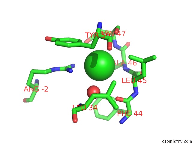
Mono view
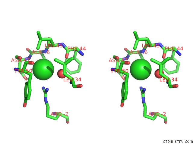
Stereo pair view
Mono view
Stereo pair view
A full contact list of Chlorine with other atoms in the Cl binding
site number 1 of The Structure of Lpxd From Pseudomonas Aeruginosa at 1.3 A Resolution within 5.0Å range:
|
Reference:
J.Badger,
B.Chie-Leon,
C.Logan,
V.Sridhar,
B.Sankaran,
P.H.Zwart,
V.Nienaber.
The Structure of Lpxd From Pseudomonas Aeruginosa at 1.3 A Resolution. Acta Crystallogr.,Sect.F V. 67 749 2011.
ISSN: ESSN 1744-3091
PubMed: 21795786
DOI: 10.1107/S1744309111018811
Page generated: Fri Jul 11 09:10:40 2025
ISSN: ESSN 1744-3091
PubMed: 21795786
DOI: 10.1107/S1744309111018811
Last articles
Fe in 6G7MFe in 6G74
Fe in 6G7Q
Fe in 6G7P
Fe in 6G7N
Fe in 6G71
Fe in 6G2J
Fe in 6G5Q
Fe in 6G5T
Fe in 6G5O