Chlorine »
PDB 3q18-3qck »
3q2m »
Chlorine in PDB 3q2m: Crystal Structure of Spectinomycin Phosphotransferase, Aph(9)-Ia, Protein Kinase Inhibitor Cki-7 Complex
Protein crystallography data
The structure of Crystal Structure of Spectinomycin Phosphotransferase, Aph(9)-Ia, Protein Kinase Inhibitor Cki-7 Complex, PDB code: 3q2m
was solved by
A.M.Berghuis,
D.H.Fong,
B.Xiong,
J.Hwang,
with X-Ray Crystallography technique. A brief refinement statistics is given in the table below:
| Resolution Low / High (Å) | 31.32 / 2.90 |
| Space group | P 31 2 1 |
| Cell size a, b, c (Å), α, β, γ (°) | 74.300, 74.300, 137.024, 90.00, 90.00, 120.00 |
| R / Rfree (%) | 22.1 / 27.9 |
Other elements in 3q2m:
The structure of Crystal Structure of Spectinomycin Phosphotransferase, Aph(9)-Ia, Protein Kinase Inhibitor Cki-7 Complex also contains other interesting chemical elements:
| Nickel | (Ni) | 1 atom |
Chlorine Binding Sites:
The binding sites of Chlorine atom in the Crystal Structure of Spectinomycin Phosphotransferase, Aph(9)-Ia, Protein Kinase Inhibitor Cki-7 Complex
(pdb code 3q2m). This binding sites where shown within
5.0 Angstroms radius around Chlorine atom.
In total only one binding site of Chlorine was determined in the Crystal Structure of Spectinomycin Phosphotransferase, Aph(9)-Ia, Protein Kinase Inhibitor Cki-7 Complex, PDB code: 3q2m:
In total only one binding site of Chlorine was determined in the Crystal Structure of Spectinomycin Phosphotransferase, Aph(9)-Ia, Protein Kinase Inhibitor Cki-7 Complex, PDB code: 3q2m:
Chlorine binding site 1 out of 1 in 3q2m
Go back to
Chlorine binding site 1 out
of 1 in the Crystal Structure of Spectinomycin Phosphotransferase, Aph(9)-Ia, Protein Kinase Inhibitor Cki-7 Complex
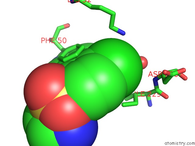
Mono view
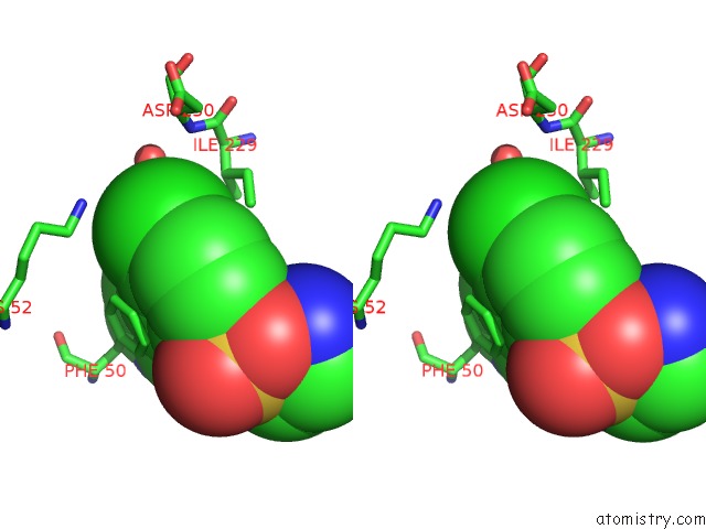
Stereo pair view
Mono view
Stereo pair view
A full contact list of Chlorine with other atoms in the Cl binding
site number 1 of Crystal Structure of Spectinomycin Phosphotransferase, Aph(9)-Ia, Protein Kinase Inhibitor Cki-7 Complex within 5.0Å range:
|
Reference:
D.H.Fong,
B.Xiong,
J.Hwang,
A.M.Berghuis.
Crystal Structures of Two Aminoglycoside Kinases Bound with A Eukaryotic Protein Kinase Inhibitor. Plos One V. 6 19589 2011.
ISSN: ESSN 1932-6203
PubMed: 21573013
DOI: 10.1371/JOURNAL.PONE.0019589
Page generated: Fri Jul 11 09:17:08 2025
ISSN: ESSN 1932-6203
PubMed: 21573013
DOI: 10.1371/JOURNAL.PONE.0019589
Last articles
Mn in 1F1UMn in 1F1R
Mn in 1EO4
Mn in 1EN6
Mn in 1EQJ
Mn in 1EN5
Mn in 1EN4
Mn in 1E9G
Mn in 1EM1
Mn in 1ELS