Chlorine »
PDB 3qp5-3qvm »
3qp5 »
Chlorine in PDB 3qp5: Crystal Structure of Cvir Bound to Antagonist Chlorolactone (Cl)
Protein crystallography data
The structure of Crystal Structure of Cvir Bound to Antagonist Chlorolactone (Cl), PDB code: 3qp5
was solved by
G.Chen,
L.Swem,
D.Swem,
D.Stauff,
C.O'loughlin,
P.Jeffrey,
B.Bassler,
F.Hughson,
with X-Ray Crystallography technique. A brief refinement statistics is given in the table below:
| Resolution Low / High (Å) | 48.88 / 3.25 |
| Space group | H 3 |
| Cell size a, b, c (Å), α, β, γ (°) | 169.318, 169.318, 99.844, 90.00, 90.00, 120.00 |
| R / Rfree (%) | 26.3 / 30.4 |
Chlorine Binding Sites:
The binding sites of Chlorine atom in the Crystal Structure of Cvir Bound to Antagonist Chlorolactone (Cl)
(pdb code 3qp5). This binding sites where shown within
5.0 Angstroms radius around Chlorine atom.
In total 4 binding sites of Chlorine where determined in the Crystal Structure of Cvir Bound to Antagonist Chlorolactone (Cl), PDB code: 3qp5:
Jump to Chlorine binding site number: 1; 2; 3; 4;
In total 4 binding sites of Chlorine where determined in the Crystal Structure of Cvir Bound to Antagonist Chlorolactone (Cl), PDB code: 3qp5:
Jump to Chlorine binding site number: 1; 2; 3; 4;
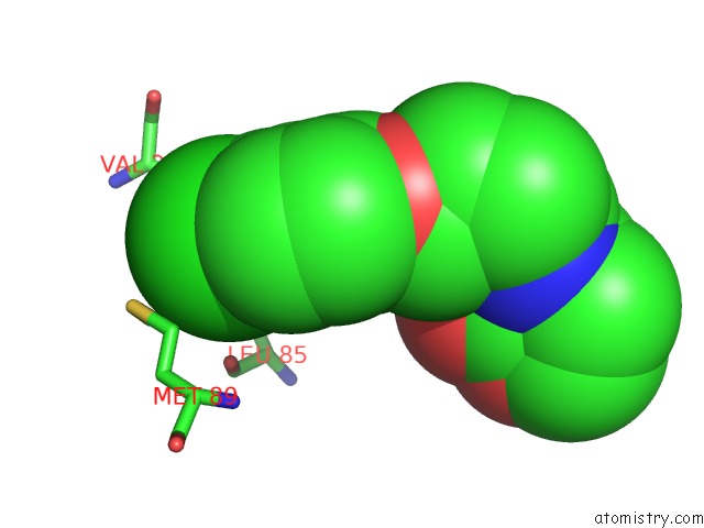
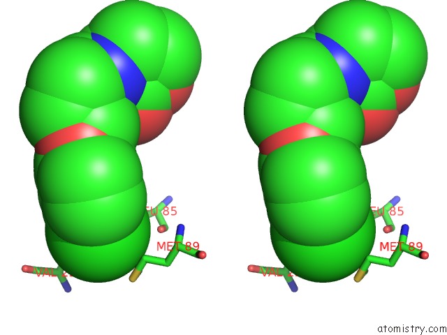
Chlorine binding site 1 out of 4 in 3qp5
Go back to
Chlorine binding site 1 out
of 4 in the Crystal Structure of Cvir Bound to Antagonist Chlorolactone (Cl)
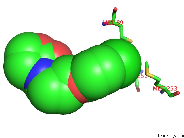
Mono view
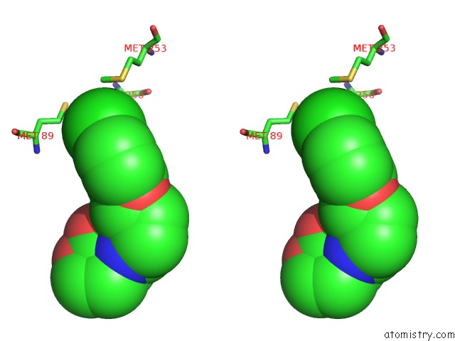
Stereo pair view
Mono view
Stereo pair view
A full contact list of Chlorine with other atoms in the Cl binding
site number 1 of Crystal Structure of Cvir Bound to Antagonist Chlorolactone (Cl) within 5.0Å range:
|
Chlorine binding site 2 out of 4 in 3qp5
Go back to
Chlorine binding site 2 out
of 4 in the Crystal Structure of Cvir Bound to Antagonist Chlorolactone (Cl)
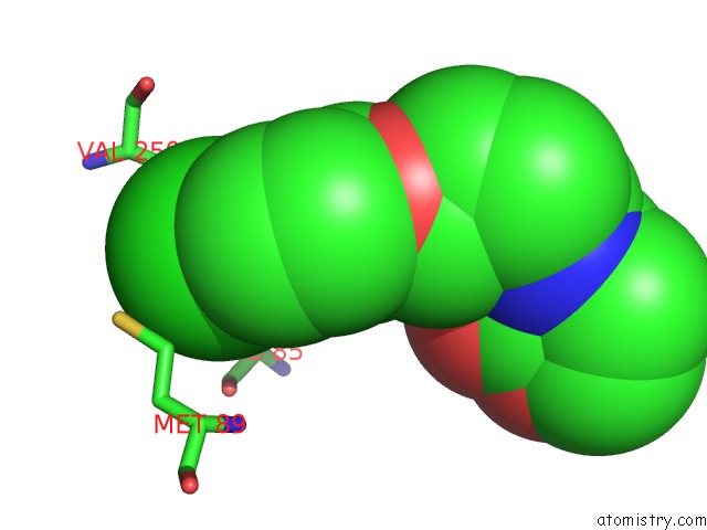
Mono view
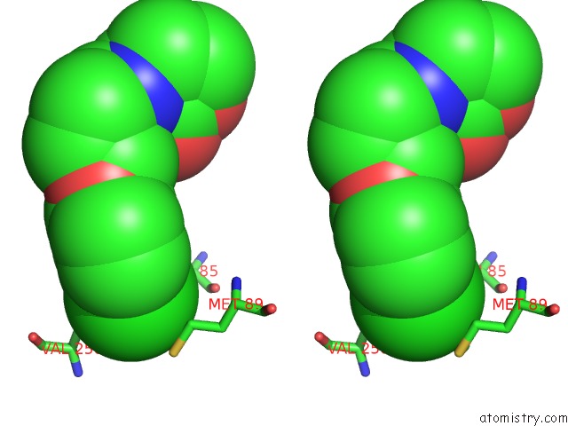
Stereo pair view
Mono view
Stereo pair view
A full contact list of Chlorine with other atoms in the Cl binding
site number 2 of Crystal Structure of Cvir Bound to Antagonist Chlorolactone (Cl) within 5.0Å range:
|
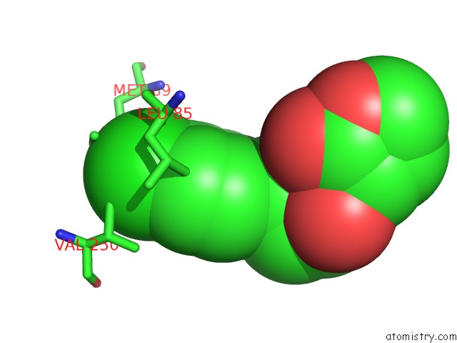
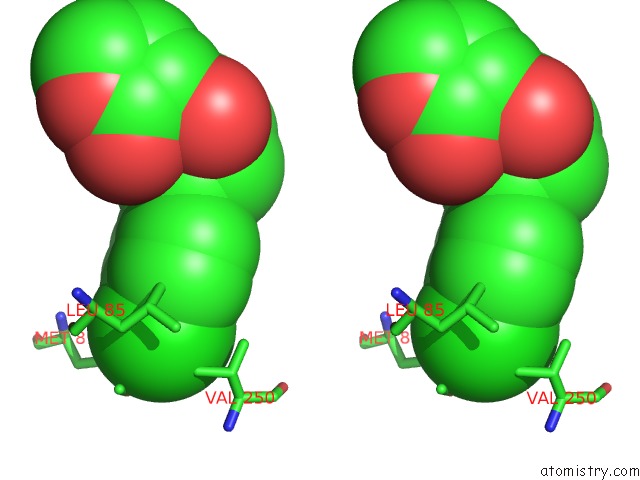
Chlorine binding site 3 out of 4 in 3qp5
Go back to
Chlorine binding site 3 out
of 4 in the Crystal Structure of Cvir Bound to Antagonist Chlorolactone (Cl)
Mono view
Stereo pair view
Mono view
Stereo pair view
A full contact list of Chlorine with other atoms in the Cl binding
site number 3 of Crystal Structure of Cvir Bound to Antagonist Chlorolactone (Cl) within 5.0Å range:
|
Chlorine binding site 4 out of 4 in 3qp5
Go back to
Chlorine binding site 4 out
of 4 in the Crystal Structure of Cvir Bound to Antagonist Chlorolactone (Cl)
Mono view
Stereo pair view
Mono view
Stereo pair view
A full contact list of Chlorine with other atoms in the Cl binding
site number 4 of Crystal Structure of Cvir Bound to Antagonist Chlorolactone (Cl) within 5.0Å range:
|
Reference:
G.Chen,
L.R.Swem,
D.L.Swem,
D.L.Stauff,
C.T.O'loughlin,
P.D.Jeffrey,
B.L.Bassler,
F.M.Hughson.
A Strategy For Antagonizing Quorum Sensing. Mol.Cell V. 42 199 2011.
ISSN: ISSN 1097-2765
PubMed: 21504831
DOI: 10.1016/J.MOLCEL.2011.04.003
Page generated: Fri Jul 11 09:29:05 2025
ISSN: ISSN 1097-2765
PubMed: 21504831
DOI: 10.1016/J.MOLCEL.2011.04.003
Last articles
Mg in 6ZZ6Mg in 6ZYM
Mg in 6ZY9
Mg in 6ZY4
Mg in 6ZVH
Mg in 6ZXQ
Mg in 6ZXM
Mg in 6ZXA
Mg in 6ZXC
Mg in 6ZXF