Chlorine »
PDB 3rmn-3ruu »
3ro8 »
Chlorine in PDB 3ro8: Crystal Structure of the Catalytic Domain of XYNA1 From Paenibacillus Sp. Jdr-2
Enzymatic activity of Crystal Structure of the Catalytic Domain of XYNA1 From Paenibacillus Sp. Jdr-2
All present enzymatic activity of Crystal Structure of the Catalytic Domain of XYNA1 From Paenibacillus Sp. Jdr-2:
3.2.1.8;
3.2.1.8;
Protein crystallography data
The structure of Crystal Structure of the Catalytic Domain of XYNA1 From Paenibacillus Sp. Jdr-2, PDB code: 3ro8
was solved by
E.Pozharski,
F.J.St John,
with X-Ray Crystallography technique. A brief refinement statistics is given in the table below:
| Resolution Low / High (Å) | 42.61 / 1.34 |
| Space group | P 1 21 1 |
| Cell size a, b, c (Å), α, β, γ (°) | 82.072, 93.172, 182.947, 90.00, 99.96, 90.00 |
| R / Rfree (%) | 14.2 / 18.5 |
Other elements in 3ro8:
The structure of Crystal Structure of the Catalytic Domain of XYNA1 From Paenibacillus Sp. Jdr-2 also contains other interesting chemical elements:
| Magnesium | (Mg) | 12 atoms |
Chlorine Binding Sites:
The binding sites of Chlorine atom in the Crystal Structure of the Catalytic Domain of XYNA1 From Paenibacillus Sp. Jdr-2
(pdb code 3ro8). This binding sites where shown within
5.0 Angstroms radius around Chlorine atom.
In total 9 binding sites of Chlorine where determined in the Crystal Structure of the Catalytic Domain of XYNA1 From Paenibacillus Sp. Jdr-2, PDB code: 3ro8:
Jump to Chlorine binding site number: 1; 2; 3; 4; 5; 6; 7; 8; 9;
In total 9 binding sites of Chlorine where determined in the Crystal Structure of the Catalytic Domain of XYNA1 From Paenibacillus Sp. Jdr-2, PDB code: 3ro8:
Jump to Chlorine binding site number: 1; 2; 3; 4; 5; 6; 7; 8; 9;
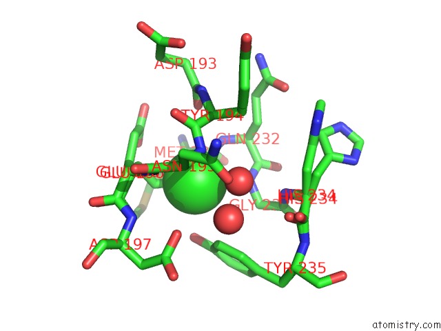
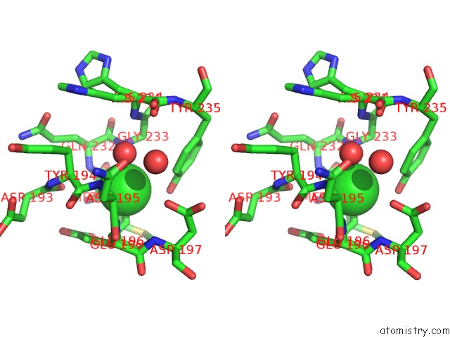
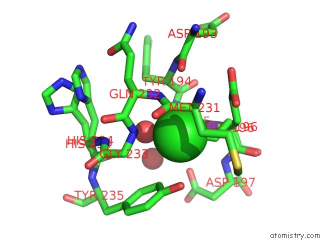
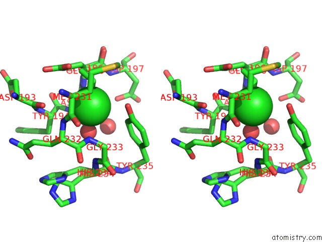
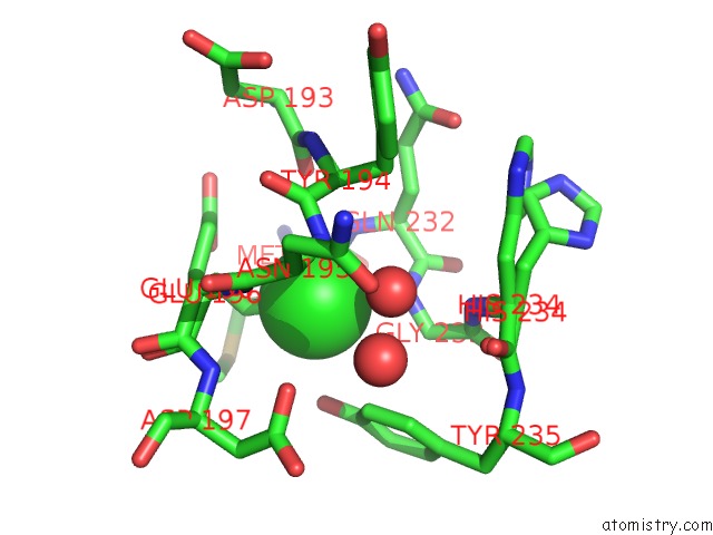
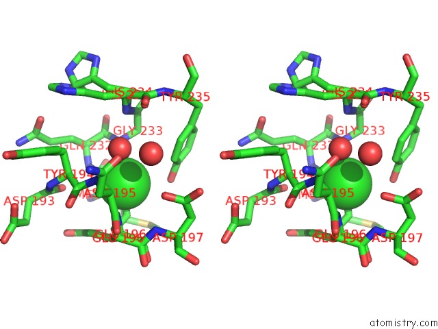
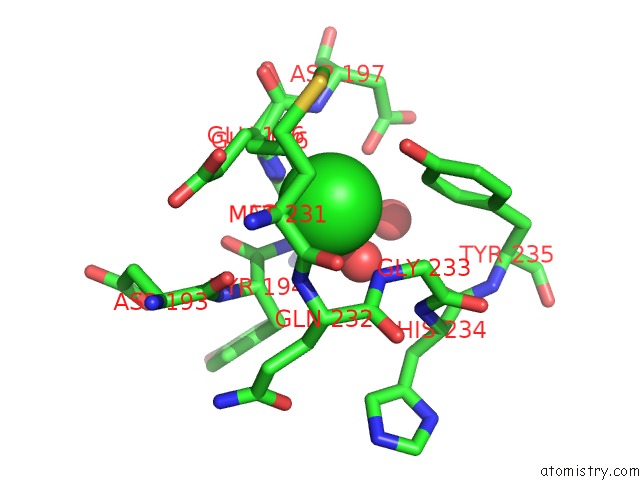
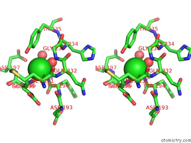
Chlorine binding site 1 out of 9 in 3ro8
Go back to
Chlorine binding site 1 out
of 9 in the Crystal Structure of the Catalytic Domain of XYNA1 From Paenibacillus Sp. Jdr-2
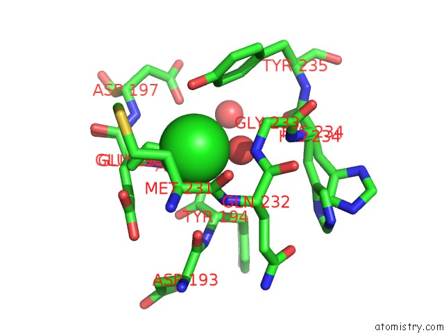
Mono view
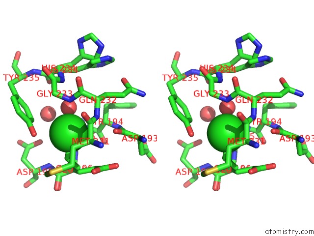
Stereo pair view
Mono view
Stereo pair view
A full contact list of Chlorine with other atoms in the Cl binding
site number 1 of Crystal Structure of the Catalytic Domain of XYNA1 From Paenibacillus Sp. Jdr-2 within 5.0Å range:
|
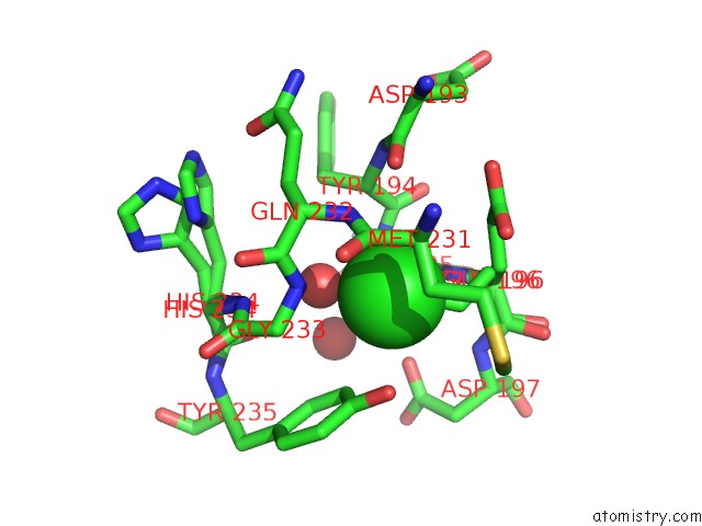
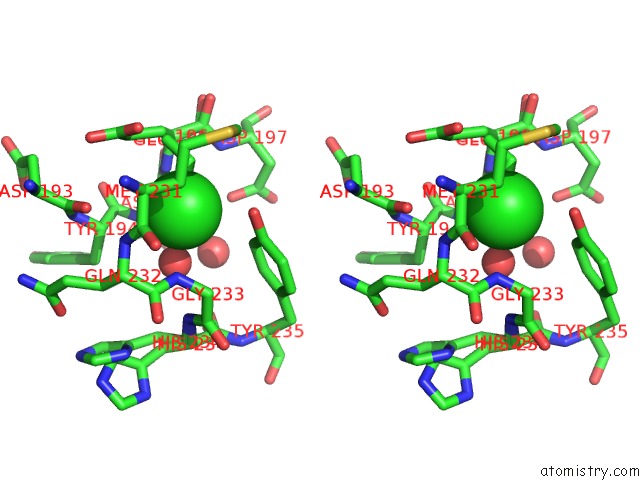
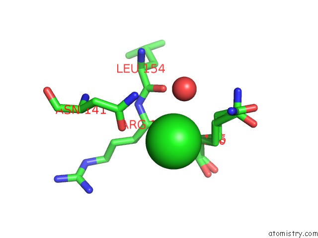
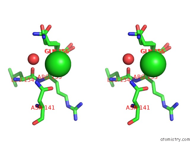
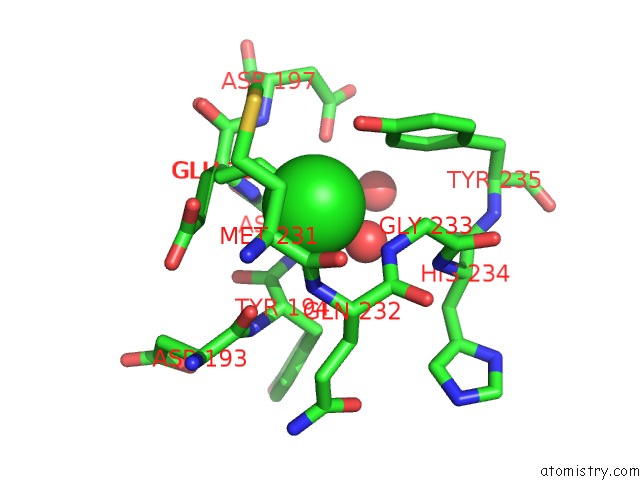
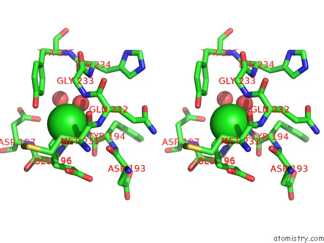
Chlorine binding site 2 out of 9 in 3ro8
Go back to
Chlorine binding site 2 out
of 9 in the Crystal Structure of the Catalytic Domain of XYNA1 From Paenibacillus Sp. Jdr-2
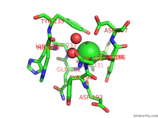
Mono view
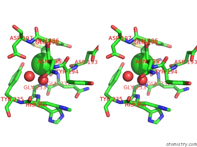
Stereo pair view
Mono view
Stereo pair view
A full contact list of Chlorine with other atoms in the Cl binding
site number 2 of Crystal Structure of the Catalytic Domain of XYNA1 From Paenibacillus Sp. Jdr-2 within 5.0Å range:
|
Chlorine binding site 3 out of 9 in 3ro8
Go back to
Chlorine binding site 3 out
of 9 in the Crystal Structure of the Catalytic Domain of XYNA1 From Paenibacillus Sp. Jdr-2
Mono view
Stereo pair view
Mono view
Stereo pair view
A full contact list of Chlorine with other atoms in the Cl binding
site number 3 of Crystal Structure of the Catalytic Domain of XYNA1 From Paenibacillus Sp. Jdr-2 within 5.0Å range:
|
Chlorine binding site 4 out of 9 in 3ro8
Go back to
Chlorine binding site 4 out
of 9 in the Crystal Structure of the Catalytic Domain of XYNA1 From Paenibacillus Sp. Jdr-2
Mono view
Stereo pair view
Mono view
Stereo pair view
A full contact list of Chlorine with other atoms in the Cl binding
site number 4 of Crystal Structure of the Catalytic Domain of XYNA1 From Paenibacillus Sp. Jdr-2 within 5.0Å range:
|
Chlorine binding site 5 out of 9 in 3ro8
Go back to
Chlorine binding site 5 out
of 9 in the Crystal Structure of the Catalytic Domain of XYNA1 From Paenibacillus Sp. Jdr-2
Mono view
Stereo pair view
Mono view
Stereo pair view
A full contact list of Chlorine with other atoms in the Cl binding
site number 5 of Crystal Structure of the Catalytic Domain of XYNA1 From Paenibacillus Sp. Jdr-2 within 5.0Å range:
|
Chlorine binding site 6 out of 9 in 3ro8
Go back to
Chlorine binding site 6 out
of 9 in the Crystal Structure of the Catalytic Domain of XYNA1 From Paenibacillus Sp. Jdr-2
Mono view
Stereo pair view
Mono view
Stereo pair view
A full contact list of Chlorine with other atoms in the Cl binding
site number 6 of Crystal Structure of the Catalytic Domain of XYNA1 From Paenibacillus Sp. Jdr-2 within 5.0Å range:
|
Chlorine binding site 7 out of 9 in 3ro8
Go back to
Chlorine binding site 7 out
of 9 in the Crystal Structure of the Catalytic Domain of XYNA1 From Paenibacillus Sp. Jdr-2
Mono view
Stereo pair view
Mono view
Stereo pair view
A full contact list of Chlorine with other atoms in the Cl binding
site number 7 of Crystal Structure of the Catalytic Domain of XYNA1 From Paenibacillus Sp. Jdr-2 within 5.0Å range:
|
Chlorine binding site 8 out of 9 in 3ro8
Go back to
Chlorine binding site 8 out
of 9 in the Crystal Structure of the Catalytic Domain of XYNA1 From Paenibacillus Sp. Jdr-2
Mono view
Stereo pair view
Mono view
Stereo pair view
A full contact list of Chlorine with other atoms in the Cl binding
site number 8 of Crystal Structure of the Catalytic Domain of XYNA1 From Paenibacillus Sp. Jdr-2 within 5.0Å range:
|
Chlorine binding site 9 out of 9 in 3ro8
Go back to
Chlorine binding site 9 out
of 9 in the Crystal Structure of the Catalytic Domain of XYNA1 From Paenibacillus Sp. Jdr-2
Mono view
Stereo pair view
Mono view
Stereo pair view
A full contact list of Chlorine with other atoms in the Cl binding
site number 9 of Crystal Structure of the Catalytic Domain of XYNA1 From Paenibacillus Sp. Jdr-2 within 5.0Å range:
|
Reference:
F.J.St John,
J.F.Preston,
E.Pozharski.
Novel Structural Features of Xylanase A1 From Paenibacillus Sp. Jdr-2. J.Struct.Biol. V. 180 303 2012.
ISSN: ISSN 1047-8477
PubMed: 23000703
DOI: 10.1016/J.JSB.2012.09.007
Page generated: Fri Jul 11 09:55:21 2025
ISSN: ISSN 1047-8477
PubMed: 23000703
DOI: 10.1016/J.JSB.2012.09.007
Last articles
Mg in 3G73Mg in 3G37
Mg in 3G6Y
Mg in 3G6X
Mg in 3G6W
Mg in 3G6V
Mg in 3G6K
Mg in 3G5A
Mg in 3G5S
Mg in 3G58