Chlorine »
PDB 3s8o-3seo »
3s8w »
Chlorine in PDB 3s8w: D2 Domain of Human IFNAR2
Protein crystallography data
The structure of D2 Domain of Human IFNAR2, PDB code: 3s8w
was solved by
C.Thomas,
K.C.Garcia,
with X-Ray Crystallography technique. A brief refinement statistics is given in the table below:
| Resolution Low / High (Å) | 19.93 / 2.60 |
| Space group | P 65 2 2 |
| Cell size a, b, c (Å), α, β, γ (°) | 82.660, 82.660, 225.330, 90.00, 90.00, 120.00 |
| R / Rfree (%) | 21.8 / 27 |
Chlorine Binding Sites:
The binding sites of Chlorine atom in the D2 Domain of Human IFNAR2
(pdb code 3s8w). This binding sites where shown within
5.0 Angstroms radius around Chlorine atom.
In total 2 binding sites of Chlorine where determined in the D2 Domain of Human IFNAR2, PDB code: 3s8w:
Jump to Chlorine binding site number: 1; 2;
In total 2 binding sites of Chlorine where determined in the D2 Domain of Human IFNAR2, PDB code: 3s8w:
Jump to Chlorine binding site number: 1; 2;
Chlorine binding site 1 out of 2 in 3s8w
Go back to
Chlorine binding site 1 out
of 2 in the D2 Domain of Human IFNAR2
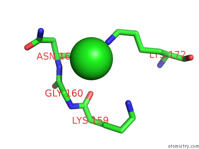
Mono view
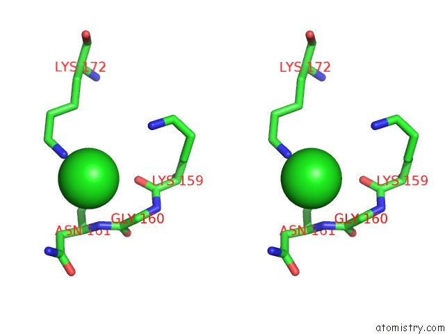
Stereo pair view
Mono view
Stereo pair view
A full contact list of Chlorine with other atoms in the Cl binding
site number 1 of D2 Domain of Human IFNAR2 within 5.0Å range:
|
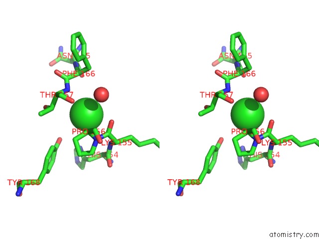
Chlorine binding site 2 out of 2 in 3s8w
Go back to
Chlorine binding site 2 out
of 2 in the D2 Domain of Human IFNAR2
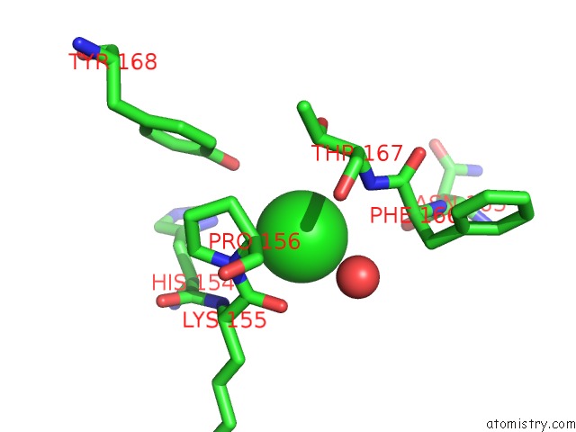
Mono view
Stereo pair view
Mono view
Stereo pair view
A full contact list of Chlorine with other atoms in the Cl binding
site number 2 of D2 Domain of Human IFNAR2 within 5.0Å range:
|
Reference:
C.Thomas,
I.Moraga,
D.Levin,
P.O.Krutzik,
Y.Podoplelova,
A.Trejo,
C.Lee,
G.Yarden,
S.E.Vleck,
J.S.Glenn,
G.P.Nolan,
J.Piehler,
G.Schreiber,
K.C.Garcia.
Structural Linkage Between Ligand Discrimination and Receptor Activation By Type I Interferons. Cell(Cambridge,Mass.) V. 146 621 2011.
ISSN: ISSN 0092-8674
PubMed: 21854986
DOI: 10.1016/J.CELL.2011.06.048
Page generated: Fri Jul 11 10:10:13 2025
ISSN: ISSN 0092-8674
PubMed: 21854986
DOI: 10.1016/J.CELL.2011.06.048
Last articles
K in 3EM2K in 3E8H
K in 3EN2
K in 3EC7
K in 3E8F
K in 3EIY
K in 3E9D
K in 3E9C
K in 3E3S
K in 3E60