Chlorine »
PDB 3ser-3smc »
3sf6 »
Chlorine in PDB 3sf6: Crystal Structure of Glutaryl-Coa Dehydrogenase From Mycobacterium Smegmatis
Enzymatic activity of Crystal Structure of Glutaryl-Coa Dehydrogenase From Mycobacterium Smegmatis
All present enzymatic activity of Crystal Structure of Glutaryl-Coa Dehydrogenase From Mycobacterium Smegmatis:
1.3.99.7;
1.3.99.7;
Protein crystallography data
The structure of Crystal Structure of Glutaryl-Coa Dehydrogenase From Mycobacterium Smegmatis, PDB code: 3sf6
was solved by
Seattle Structural Genomics Center For Infectious Disease (Ssgcid),
with X-Ray Crystallography technique. A brief refinement statistics is given in the table below:
| Resolution Low / High (Å) | 44.57 / 1.70 |
| Space group | I 41 2 2 |
| Cell size a, b, c (Å), α, β, γ (°) | 130.000, 130.000, 101.930, 90.00, 90.00, 90.00 |
| R / Rfree (%) | 12.8 / 15.2 |
Chlorine Binding Sites:
The binding sites of Chlorine atom in the Crystal Structure of Glutaryl-Coa Dehydrogenase From Mycobacterium Smegmatis
(pdb code 3sf6). This binding sites where shown within
5.0 Angstroms radius around Chlorine atom.
In total 2 binding sites of Chlorine where determined in the Crystal Structure of Glutaryl-Coa Dehydrogenase From Mycobacterium Smegmatis, PDB code: 3sf6:
Jump to Chlorine binding site number: 1; 2;
In total 2 binding sites of Chlorine where determined in the Crystal Structure of Glutaryl-Coa Dehydrogenase From Mycobacterium Smegmatis, PDB code: 3sf6:
Jump to Chlorine binding site number: 1; 2;
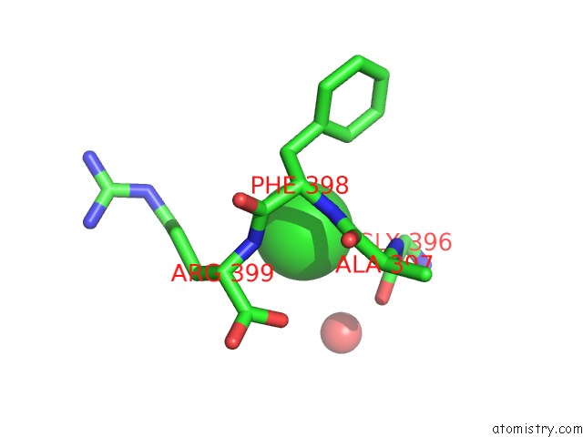
Chlorine binding site 1 out of 2 in 3sf6
Go back to
Chlorine binding site 1 out
of 2 in the Crystal Structure of Glutaryl-Coa Dehydrogenase From Mycobacterium Smegmatis

Mono view

Stereo pair view
Mono view
Stereo pair view
A full contact list of Chlorine with other atoms in the Cl binding
site number 1 of Crystal Structure of Glutaryl-Coa Dehydrogenase From Mycobacterium Smegmatis within 5.0Å range:
|
Chlorine binding site 2 out of 2 in 3sf6
Go back to
Chlorine binding site 2 out
of 2 in the Crystal Structure of Glutaryl-Coa Dehydrogenase From Mycobacterium Smegmatis
Mono view

Stereo pair view
Mono view
Stereo pair view
A full contact list of Chlorine with other atoms in the Cl binding
site number 2 of Crystal Structure of Glutaryl-Coa Dehydrogenase From Mycobacterium Smegmatis within 5.0Å range:
|
Reference:
L.Baugh,
I.Phan,
D.W.Begley,
M.C.Clifton,
B.Armour,
D.M.Dranow,
B.M.Taylor,
M.M.Muruthi,
J.Abendroth,
J.W.Fairman,
D.Fox,
S.H.Dieterich,
B.L.Staker,
A.S.Gardberg,
R.Choi,
S.N.Hewitt,
A.J.Napuli,
J.Myers,
L.K.Barrett,
Y.Zhang,
M.Ferrell,
E.Mundt,
K.Thompkins,
N.Tran,
S.Lyons-Abbott,
A.Abramov,
A.Sekar,
D.Serbzhinskiy,
D.Lorimer,
G.W.Buchko,
R.Stacy,
L.J.Stewart,
T.E.Edwards,
W.C.Van Voorhis,
P.J.Myler.
Increasing the Structural Coverage of Tuberculosis Drug Targets. Tuberculosis (Edinb) V. 95 142 2015.
ISSN: ISSN 1472-9792
PubMed: 25613812
DOI: 10.1016/J.TUBE.2014.12.003
Page generated: Fri Jul 11 10:17:34 2025
ISSN: ISSN 1472-9792
PubMed: 25613812
DOI: 10.1016/J.TUBE.2014.12.003
Last articles
Fe in 2YXOFe in 2YRS
Fe in 2YXC
Fe in 2YNM
Fe in 2YVJ
Fe in 2YP1
Fe in 2YU2
Fe in 2YU1
Fe in 2YQB
Fe in 2YOO