Chlorine »
PDB 3sml-3ste »
3sol »
Chlorine in PDB 3sol: Crystal Structure of the Type 2 Secretion System Pilotin Gsps
Protein crystallography data
The structure of Crystal Structure of the Type 2 Secretion System Pilotin Gsps, PDB code: 3sol
was solved by
K.V.Korotkov,
W.G.J.Hol,
with X-Ray Crystallography technique. A brief refinement statistics is given in the table below:
| Resolution Low / High (Å) | 47.26 / 1.90 |
| Space group | P 61 2 2 |
| Cell size a, b, c (Å), α, β, γ (°) | 73.350, 73.350, 70.730, 90.00, 90.00, 120.00 |
| R / Rfree (%) | 19 / 22.2 |
Chlorine Binding Sites:
The binding sites of Chlorine atom in the Crystal Structure of the Type 2 Secretion System Pilotin Gsps
(pdb code 3sol). This binding sites where shown within
5.0 Angstroms radius around Chlorine atom.
In total 4 binding sites of Chlorine where determined in the Crystal Structure of the Type 2 Secretion System Pilotin Gsps, PDB code: 3sol:
Jump to Chlorine binding site number: 1; 2; 3; 4;
In total 4 binding sites of Chlorine where determined in the Crystal Structure of the Type 2 Secretion System Pilotin Gsps, PDB code: 3sol:
Jump to Chlorine binding site number: 1; 2; 3; 4;
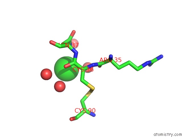
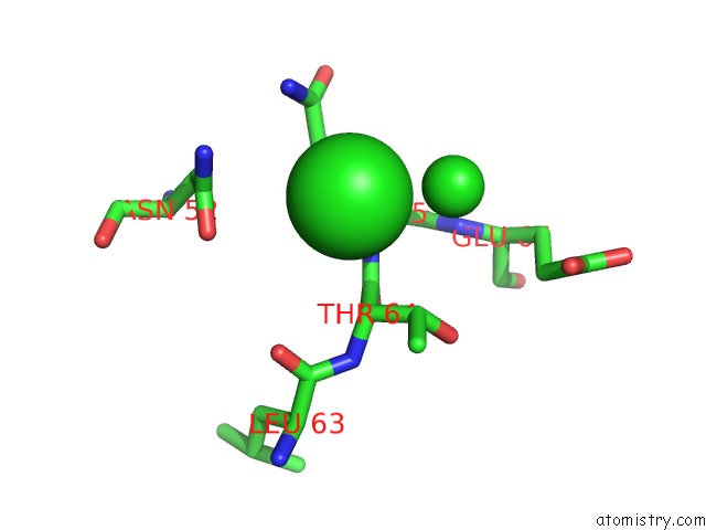
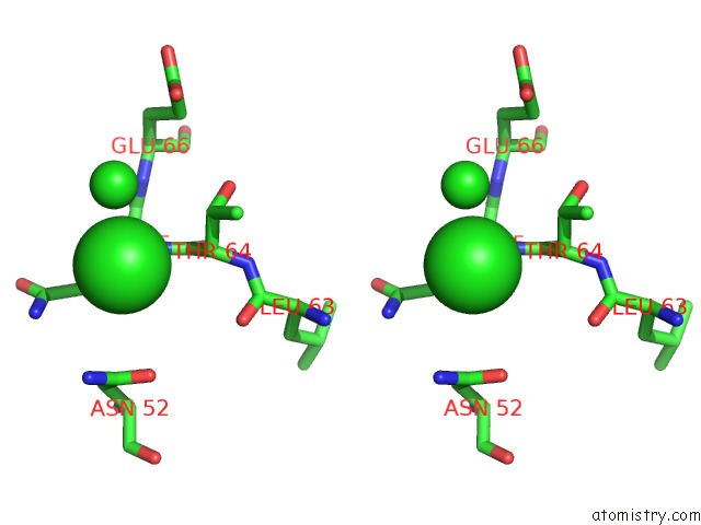
Chlorine binding site 1 out of 4 in 3sol
Go back to
Chlorine binding site 1 out
of 4 in the Crystal Structure of the Type 2 Secretion System Pilotin Gsps
Mono view
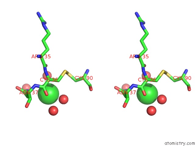
Stereo pair view
Mono view
Stereo pair view
A full contact list of Chlorine with other atoms in the Cl binding
site number 1 of Crystal Structure of the Type 2 Secretion System Pilotin Gsps within 5.0Å range:
|
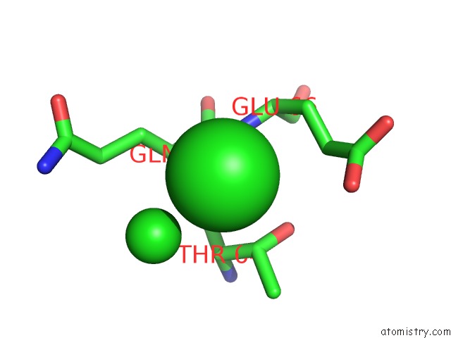
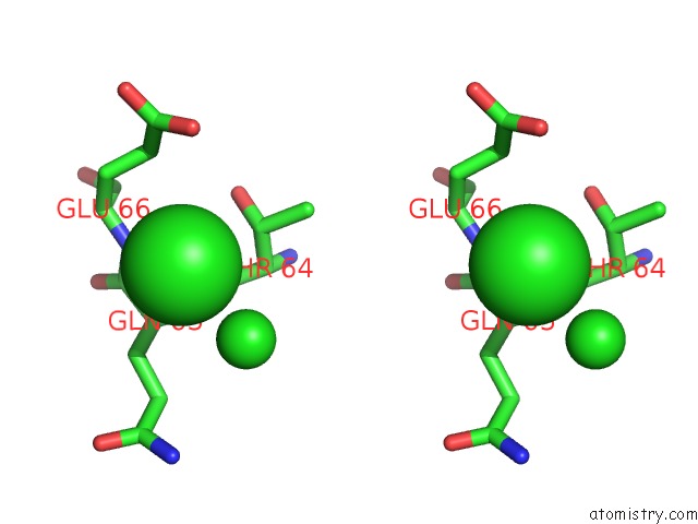
Chlorine binding site 2 out of 4 in 3sol
Go back to
Chlorine binding site 2 out
of 4 in the Crystal Structure of the Type 2 Secretion System Pilotin Gsps
Mono view
Stereo pair view
Mono view
Stereo pair view
A full contact list of Chlorine with other atoms in the Cl binding
site number 2 of Crystal Structure of the Type 2 Secretion System Pilotin Gsps within 5.0Å range:
|
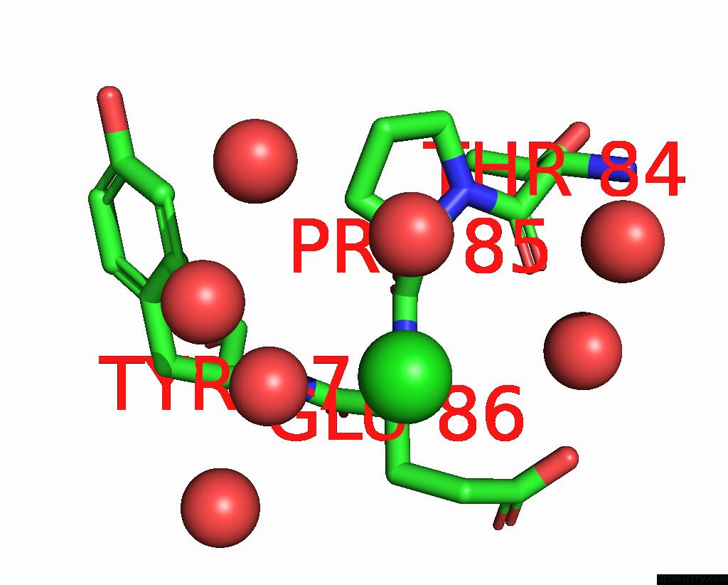
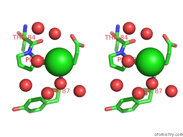
Chlorine binding site 3 out of 4 in 3sol
Go back to
Chlorine binding site 3 out
of 4 in the Crystal Structure of the Type 2 Secretion System Pilotin Gsps
Mono view
Stereo pair view
Mono view
Stereo pair view
A full contact list of Chlorine with other atoms in the Cl binding
site number 3 of Crystal Structure of the Type 2 Secretion System Pilotin Gsps within 5.0Å range:
|
Chlorine binding site 4 out of 4 in 3sol
Go back to
Chlorine binding site 4 out
of 4 in the Crystal Structure of the Type 2 Secretion System Pilotin Gsps
Mono view
Stereo pair view
Mono view
Stereo pair view
A full contact list of Chlorine with other atoms in the Cl binding
site number 4 of Crystal Structure of the Type 2 Secretion System Pilotin Gsps within 5.0Å range:
|
Reference:
K.V.Korotkov,
W.G.Hol.
Crystal Structure of the Pilotin From the Enterohemorrhagic Escherichia Coli Type II Secretion System. J.Struct.Biol. V. 182 186 2013.
ISSN: ISSN 1047-8477
PubMed: 23458689
DOI: 10.1016/J.JSB.2013.02.013
Page generated: Fri Jul 11 10:25:16 2025
ISSN: ISSN 1047-8477
PubMed: 23458689
DOI: 10.1016/J.JSB.2013.02.013
Last articles
Mg in 6ZZ6Mg in 6ZYM
Mg in 6ZY9
Mg in 6ZY4
Mg in 6ZVH
Mg in 6ZXQ
Mg in 6ZXM
Mg in 6ZXA
Mg in 6ZXC
Mg in 6ZXF