Chlorine »
PDB 3sml-3ste »
3ss3 »
Chlorine in PDB 3ss3: Crystal Structure of Mouse Glutaminase C, Ligand-Free Form
Enzymatic activity of Crystal Structure of Mouse Glutaminase C, Ligand-Free Form
All present enzymatic activity of Crystal Structure of Mouse Glutaminase C, Ligand-Free Form:
3.5.1.2;
3.5.1.2;
Protein crystallography data
The structure of Crystal Structure of Mouse Glutaminase C, Ligand-Free Form, PDB code: 3ss3
was solved by
A.L.B.Ambrosio,
S.M.G.Dias,
R.A.Cerione,
with X-Ray Crystallography technique. A brief refinement statistics is given in the table below:
| Resolution Low / High (Å) | 19.93 / 2.42 |
| Space group | P 21 21 21 |
| Cell size a, b, c (Å), α, β, γ (°) | 99.260, 138.810, 179.480, 90.00, 90.00, 90.00 |
| R / Rfree (%) | 19.5 / 25 |
Chlorine Binding Sites:
The binding sites of Chlorine atom in the Crystal Structure of Mouse Glutaminase C, Ligand-Free Form
(pdb code 3ss3). This binding sites where shown within
5.0 Angstroms radius around Chlorine atom.
In total 4 binding sites of Chlorine where determined in the Crystal Structure of Mouse Glutaminase C, Ligand-Free Form, PDB code: 3ss3:
Jump to Chlorine binding site number: 1; 2; 3; 4;
In total 4 binding sites of Chlorine where determined in the Crystal Structure of Mouse Glutaminase C, Ligand-Free Form, PDB code: 3ss3:
Jump to Chlorine binding site number: 1; 2; 3; 4;
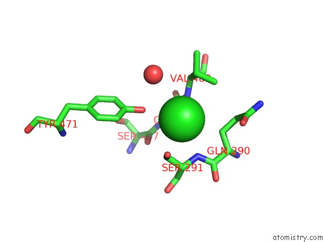
Chlorine binding site 1 out of 4 in 3ss3
Go back to
Chlorine binding site 1 out
of 4 in the Crystal Structure of Mouse Glutaminase C, Ligand-Free Form

Mono view
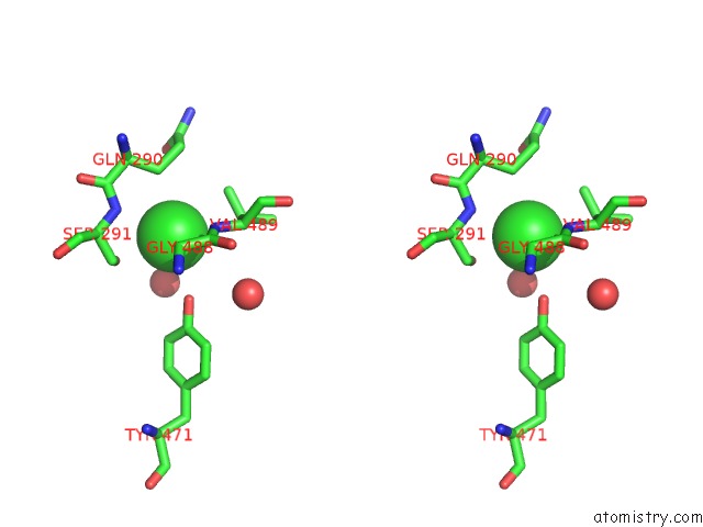
Stereo pair view
Mono view
Stereo pair view
A full contact list of Chlorine with other atoms in the Cl binding
site number 1 of Crystal Structure of Mouse Glutaminase C, Ligand-Free Form within 5.0Å range:
|
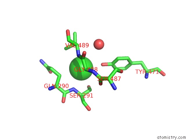
Chlorine binding site 2 out of 4 in 3ss3
Go back to
Chlorine binding site 2 out
of 4 in the Crystal Structure of Mouse Glutaminase C, Ligand-Free Form
Mono view

Stereo pair view
Mono view
Stereo pair view
A full contact list of Chlorine with other atoms in the Cl binding
site number 2 of Crystal Structure of Mouse Glutaminase C, Ligand-Free Form within 5.0Å range:
|
Chlorine binding site 3 out of 4 in 3ss3
Go back to
Chlorine binding site 3 out
of 4 in the Crystal Structure of Mouse Glutaminase C, Ligand-Free Form
Mono view

Stereo pair view
Mono view
Stereo pair view
A full contact list of Chlorine with other atoms in the Cl binding
site number 3 of Crystal Structure of Mouse Glutaminase C, Ligand-Free Form within 5.0Å range:
|
Chlorine binding site 4 out of 4 in 3ss3
Go back to
Chlorine binding site 4 out
of 4 in the Crystal Structure of Mouse Glutaminase C, Ligand-Free Form

Mono view

Stereo pair view
Mono view
Stereo pair view
A full contact list of Chlorine with other atoms in the Cl binding
site number 4 of Crystal Structure of Mouse Glutaminase C, Ligand-Free Form within 5.0Å range:
|
Reference:
A.Cassago,
A.P.Ferreira,
I.M.Ferreira,
C.Fornezari,
E.R.Gomes,
K.S.Greene,
H.M.Pereira,
R.C.Garratt,
S.M.Dias,
A.L.Ambrosio.
Mitochondrial Localization and Structure-Based Phosphate Activation Mechanism of Glutaminase C with Implications For Cancer Metabolism. Proc.Natl.Acad.Sci.Usa V. 109 1092 2012.
ISSN: ISSN 0027-8424
PubMed: 22228304
DOI: 10.1073/PNAS.1112495109
Page generated: Fri Jul 11 10:26:58 2025
ISSN: ISSN 0027-8424
PubMed: 22228304
DOI: 10.1073/PNAS.1112495109
Last articles
Mg in 6ZZ6Mg in 6ZYM
Mg in 6ZY9
Mg in 6ZY4
Mg in 6ZVH
Mg in 6ZXQ
Mg in 6ZXM
Mg in 6ZXA
Mg in 6ZXC
Mg in 6ZXF