Chlorine »
PDB 3th4-3tnn »
3thd »
Chlorine in PDB 3thd: Crystal Structure of Human Beta-Galactosidase in Complex with 1- Deoxygalactonojirimycin
Enzymatic activity of Crystal Structure of Human Beta-Galactosidase in Complex with 1- Deoxygalactonojirimycin
All present enzymatic activity of Crystal Structure of Human Beta-Galactosidase in Complex with 1- Deoxygalactonojirimycin:
3.2.1.23;
3.2.1.23;
Protein crystallography data
The structure of Crystal Structure of Human Beta-Galactosidase in Complex with 1- Deoxygalactonojirimycin, PDB code: 3thd
was solved by
U.Ohto,
T.Shimizu,
with X-Ray Crystallography technique. A brief refinement statistics is given in the table below:
| Resolution Low / High (Å) | 29.29 / 1.79 |
| Space group | P 1 21 1 |
| Cell size a, b, c (Å), α, β, γ (°) | 94.792, 115.996, 140.327, 90.00, 92.22, 90.00 |
| R / Rfree (%) | 19.8 / 22.8 |
Chlorine Binding Sites:
The binding sites of Chlorine atom in the Crystal Structure of Human Beta-Galactosidase in Complex with 1- Deoxygalactonojirimycin
(pdb code 3thd). This binding sites where shown within
5.0 Angstroms radius around Chlorine atom.
In total 4 binding sites of Chlorine where determined in the Crystal Structure of Human Beta-Galactosidase in Complex with 1- Deoxygalactonojirimycin, PDB code: 3thd:
Jump to Chlorine binding site number: 1; 2; 3; 4;
In total 4 binding sites of Chlorine where determined in the Crystal Structure of Human Beta-Galactosidase in Complex with 1- Deoxygalactonojirimycin, PDB code: 3thd:
Jump to Chlorine binding site number: 1; 2; 3; 4;
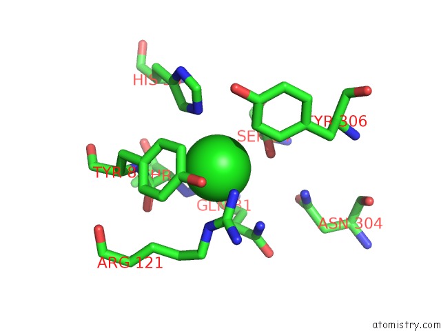
Chlorine binding site 1 out of 4 in 3thd
Go back to
Chlorine binding site 1 out
of 4 in the Crystal Structure of Human Beta-Galactosidase in Complex with 1- Deoxygalactonojirimycin
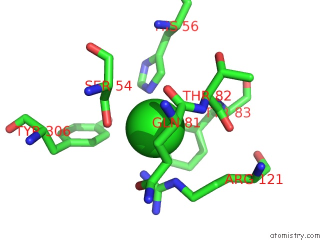
Mono view
Stereo pair view
Mono view
Stereo pair view
A full contact list of Chlorine with other atoms in the Cl binding
site number 1 of Crystal Structure of Human Beta-Galactosidase in Complex with 1- Deoxygalactonojirimycin within 5.0Å range:
|
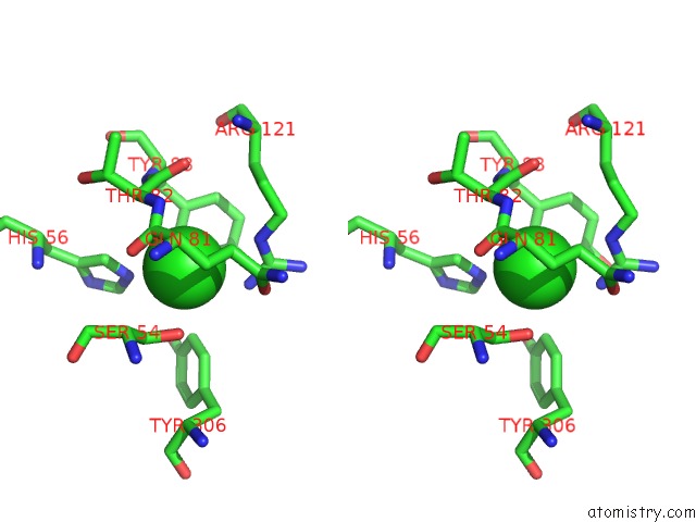
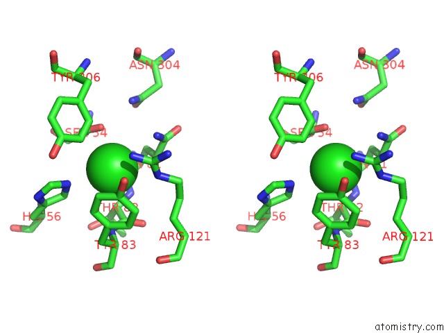
Chlorine binding site 2 out of 4 in 3thd
Go back to
Chlorine binding site 2 out
of 4 in the Crystal Structure of Human Beta-Galactosidase in Complex with 1- Deoxygalactonojirimycin
Mono view
Stereo pair view
Mono view
Stereo pair view
A full contact list of Chlorine with other atoms in the Cl binding
site number 2 of Crystal Structure of Human Beta-Galactosidase in Complex with 1- Deoxygalactonojirimycin within 5.0Å range:
|
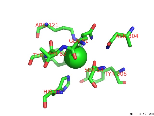
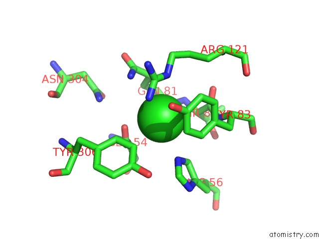
Chlorine binding site 3 out of 4 in 3thd
Go back to
Chlorine binding site 3 out
of 4 in the Crystal Structure of Human Beta-Galactosidase in Complex with 1- Deoxygalactonojirimycin
Mono view
Stereo pair view
Mono view
Stereo pair view
A full contact list of Chlorine with other atoms in the Cl binding
site number 3 of Crystal Structure of Human Beta-Galactosidase in Complex with 1- Deoxygalactonojirimycin within 5.0Å range:
|
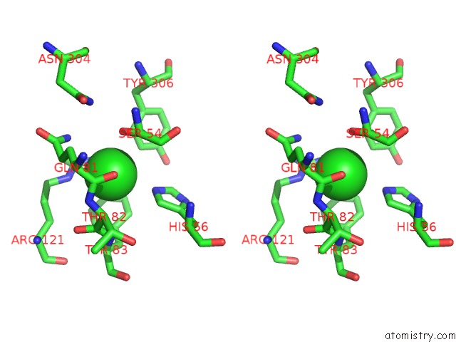
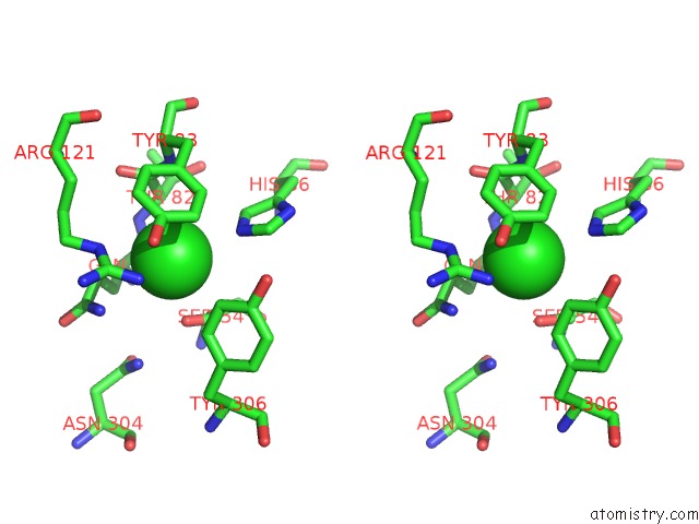
Chlorine binding site 4 out of 4 in 3thd
Go back to
Chlorine binding site 4 out
of 4 in the Crystal Structure of Human Beta-Galactosidase in Complex with 1- Deoxygalactonojirimycin
Mono view
Stereo pair view
Mono view
Stereo pair view
A full contact list of Chlorine with other atoms in the Cl binding
site number 4 of Crystal Structure of Human Beta-Galactosidase in Complex with 1- Deoxygalactonojirimycin within 5.0Å range:
|
Reference:
U.Ohto,
K.Usui,
T.Ochi,
K.Yuki,
Y.Satow,
T.Shimizu.
Crystal Structure of Human Beta-Galactosidase: Structural Basis of GM1 Gangliosidosis and Morquio B Diseases J.Biol.Chem. V. 287 1801 2012.
ISSN: ISSN 0021-9258
PubMed: 22128166
DOI: 10.1074/JBC.M111.293795
Page generated: Fri Jul 11 10:44:07 2025
ISSN: ISSN 0021-9258
PubMed: 22128166
DOI: 10.1074/JBC.M111.293795
Last articles
Mg in 2X0QMg in 2X03
Mg in 2WZG
Mg in 2WZD
Mg in 2WZC
Mg in 2WZB
Mg in 2WZ8
Mg in 2WX5
Mg in 2WWR
Mg in 2WW8