Chlorine »
PDB 3th4-3tnn »
3tja »
Chlorine in PDB 3tja: Crystal Structure of Helicobacter Pylori Uree in the Apo Form
Protein crystallography data
The structure of Crystal Structure of Helicobacter Pylori Uree in the Apo Form, PDB code: 3tja
was solved by
K.Banaszak,
M.Bellucci,
B.Zambelli,
W.R.Rypniewski,
S.Ciurli,
with X-Ray Crystallography technique. A brief refinement statistics is given in the table below:
| Resolution Low / High (Å) | 19.70 / 2.00 |
| Space group | P 21 21 21 |
| Cell size a, b, c (Å), α, β, γ (°) | 68.999, 70.468, 123.340, 90.00, 90.00, 90.00 |
| R / Rfree (%) | 20.8 / 26.7 |
Chlorine Binding Sites:
The binding sites of Chlorine atom in the Crystal Structure of Helicobacter Pylori Uree in the Apo Form
(pdb code 3tja). This binding sites where shown within
5.0 Angstroms radius around Chlorine atom.
In total only one binding site of Chlorine was determined in the Crystal Structure of Helicobacter Pylori Uree in the Apo Form, PDB code: 3tja:
In total only one binding site of Chlorine was determined in the Crystal Structure of Helicobacter Pylori Uree in the Apo Form, PDB code: 3tja:
Chlorine binding site 1 out of 1 in 3tja
Go back to
Chlorine binding site 1 out
of 1 in the Crystal Structure of Helicobacter Pylori Uree in the Apo Form
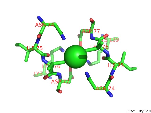
Mono view
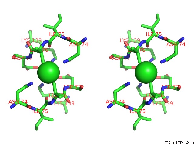
Stereo pair view
Mono view
Stereo pair view
A full contact list of Chlorine with other atoms in the Cl binding
site number 1 of Crystal Structure of Helicobacter Pylori Uree in the Apo Form within 5.0Å range:
|
Reference:
K.Banaszak,
V.Martin-Diaconescu,
M.Bellucci,
B.Zambelli,
W.Rypniewski,
M.J.Maroney,
S.Ciurli.
Crystallographic and X-Ray Absorption Spectroscopic Characterization of Helicobacter Pylori Uree Bound to NI2+ and ZN2+ Reveals A Role For the Disordered C-Terminal Arm in Metal Trafficking. Biochem.J. V. 441 1017 2012.
ISSN: ISSN 0264-6021
PubMed: 22010876
DOI: 10.1042/BJ20111659
Page generated: Fri Jul 11 10:46:45 2025
ISSN: ISSN 0264-6021
PubMed: 22010876
DOI: 10.1042/BJ20111659
Last articles
Mg in 3U05Mg in 3TYZ
Mg in 3TZ5
Mg in 3TZ4
Mg in 3TXA
Mg in 3TY7
Mg in 3TX9
Mg in 3TXZ
Mg in 3TWP
Mg in 3TWB