Chlorine »
PDB 3tno-3txf »
3tug »
Chlorine in PDB 3tug: Crystal Structure of the Hect Domain of Itch E3 Ubiquitin Ligase
Protein crystallography data
The structure of Crystal Structure of the Hect Domain of Itch E3 Ubiquitin Ligase, PDB code: 3tug
was solved by
A.Dong,
E.Dobrovetsky,
S.Xue,
C.Butler,
A.Wernimont,
J.R.Walker,
W.Tempel,
S.Dhe-Paganon,
C.H.Arrowsmith,
A.M.Edwards,
C.Bountra,
Y.Tong,
Structuralgenomics Consortium (Sgc),
with X-Ray Crystallography technique. A brief refinement statistics is given in the table below:
| Resolution Low / High (Å) | 38.74 / 2.27 |
| Space group | P 31 2 1 |
| Cell size a, b, c (Å), α, β, γ (°) | 82.779, 82.779, 109.943, 90.00, 90.00, 120.00 |
| R / Rfree (%) | 19.6 / 25.6 |
Chlorine Binding Sites:
The binding sites of Chlorine atom in the Crystal Structure of the Hect Domain of Itch E3 Ubiquitin Ligase
(pdb code 3tug). This binding sites where shown within
5.0 Angstroms radius around Chlorine atom.
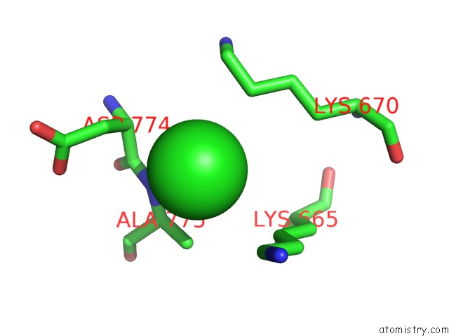
In total only one binding site of Chlorine was determined in the Crystal Structure of the Hect Domain of Itch E3 Ubiquitin Ligase, PDB code: 3tug:
In total only one binding site of Chlorine was determined in the Crystal Structure of the Hect Domain of Itch E3 Ubiquitin Ligase, PDB code: 3tug:
Chlorine binding site 1 out of 1 in 3tug
Go back to
Chlorine binding site 1 out
of 1 in the Crystal Structure of the Hect Domain of Itch E3 Ubiquitin Ligase
Mono view

Stereo pair view
Mono view
Stereo pair view
A full contact list of Chlorine with other atoms in the Cl binding
site number 1 of Crystal Structure of the Hect Domain of Itch E3 Ubiquitin Ligase within 5.0Å range:
|
Reference:
E.Dobrovetsky,
A.Dong,
S.Xue,
C.Butler,
A.Wernimont,
J.R.Walker,
W.Tempel,
S.Dhe-Paganon,
C.H.Arrowsmith,
A.M.Edwards,
C.Bountra,
Y.Tong.
Crystal Structure of the Hect Domain of Itch E3 Ubiquitin Ligase To Be Published.
Page generated: Fri Jul 11 10:55:49 2025
Last articles
Zn in 3MZ8Zn in 3MZC
Zn in 3MYQ
Zn in 3MX2
Zn in 3MXW
Zn in 3MWT
Zn in 3MWM
Zn in 3MWP
Zn in 3MUP
Zn in 3MWO