Chlorine »
PDB 3u6a-3ue5 »
3u9e »
Chlorine in PDB 3u9e: The Crystal Structure of A Possible Phosphate Acetyl/Butaryl Transferase (From Listeria Monocytogenes Egd-E) in Complex with Coa.
Protein crystallography data
The structure of The Crystal Structure of A Possible Phosphate Acetyl/Butaryl Transferase (From Listeria Monocytogenes Egd-E) in Complex with Coa., PDB code: 3u9e
was solved by
K.Tan,
M.Zhou,
S.Peterson,
W.F.Anderson,
A.Joachimiak,
Center Forstructural Genomics Of Infectious Diseases (Csgid),
with X-Ray Crystallography technique. A brief refinement statistics is given in the table below:
| Resolution Low / High (Å) | 38.84 / 2.04 |
| Space group | P 1 21 1 |
| Cell size a, b, c (Å), α, β, γ (°) | 43.739, 76.727, 80.090, 90.00, 104.08, 90.00 |
| R / Rfree (%) | 18.8 / 24.2 |
Chlorine Binding Sites:
The binding sites of Chlorine atom in the The Crystal Structure of A Possible Phosphate Acetyl/Butaryl Transferase (From Listeria Monocytogenes Egd-E) in Complex with Coa.
(pdb code 3u9e). This binding sites where shown within
5.0 Angstroms radius around Chlorine atom.
In total only one binding site of Chlorine was determined in the The Crystal Structure of A Possible Phosphate Acetyl/Butaryl Transferase (From Listeria Monocytogenes Egd-E) in Complex with Coa., PDB code: 3u9e:
In total only one binding site of Chlorine was determined in the The Crystal Structure of A Possible Phosphate Acetyl/Butaryl Transferase (From Listeria Monocytogenes Egd-E) in Complex with Coa., PDB code: 3u9e:
Chlorine binding site 1 out of 1 in 3u9e
Go back to
Chlorine binding site 1 out
of 1 in the The Crystal Structure of A Possible Phosphate Acetyl/Butaryl Transferase (From Listeria Monocytogenes Egd-E) in Complex with Coa.
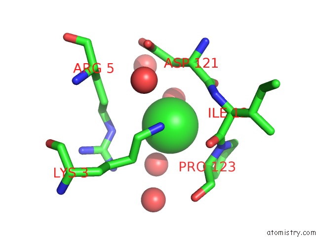
Mono view
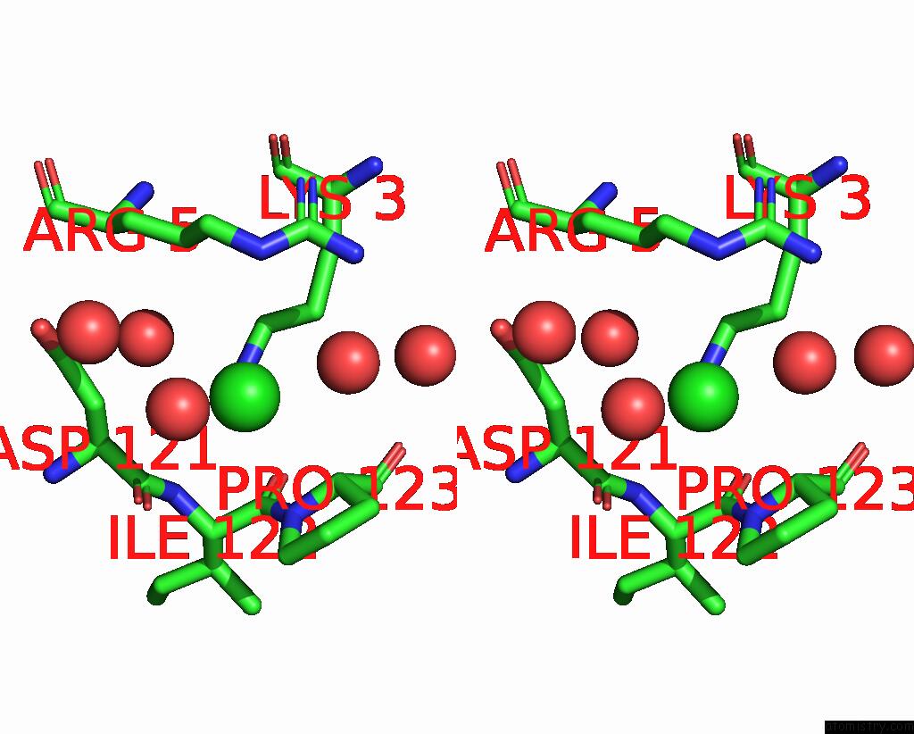
Stereo pair view
Mono view
Stereo pair view
A full contact list of Chlorine with other atoms in the Cl binding
site number 1 of The Crystal Structure of A Possible Phosphate Acetyl/Butaryl Transferase (From Listeria Monocytogenes Egd-E) in Complex with Coa. within 5.0Å range:
|
Reference:
K.Tan,
M.Zhou,
S.Peterson,
W.F.Anderson,
A.Joachimiak.
The Crystal Structure of A Possible Phosphate Acetyl/Butaryl Transferase (From Listeria Monocytogenes Egd-E) in Complex with Coa. To Be Published.
Page generated: Fri Jul 11 11:09:51 2025
Last articles
Mg in 1L8AMg in 1L8Q
Mg in 1L7N
Mg in 1L7M
Mg in 1L6Y
Mg in 1L6S
Mg in 1L5Y
Mg in 1L5U
Mg in 1L4Y
Mg in 1L3V