Chlorine »
PDB 3u6a-3ue5 »
3ubt »
Chlorine in PDB 3ubt: Crystal Structure of C71S Mutant of Dna Cytosine-5 Methyltransferase M.Haeiii Bound to Dna
Enzymatic activity of Crystal Structure of C71S Mutant of Dna Cytosine-5 Methyltransferase M.Haeiii Bound to Dna
All present enzymatic activity of Crystal Structure of C71S Mutant of Dna Cytosine-5 Methyltransferase M.Haeiii Bound to Dna:
2.1.1.37;
2.1.1.37;
Protein crystallography data
The structure of Crystal Structure of C71S Mutant of Dna Cytosine-5 Methyltransferase M.Haeiii Bound to Dna, PDB code: 3ubt
was solved by
G.L.Verdine,
A.Didovyk,
with X-Ray Crystallography technique. A brief refinement statistics is given in the table below:
| Resolution Low / High (Å) | 16.98 / 2.50 |
| Space group | P 21 21 21 |
| Cell size a, b, c (Å), α, β, γ (°) | 57.564, 129.588, 132.381, 90.00, 90.00, 90.00 |
| R / Rfree (%) | 18.2 / 22.1 |
Chlorine Binding Sites:
The binding sites of Chlorine atom in the Crystal Structure of C71S Mutant of Dna Cytosine-5 Methyltransferase M.Haeiii Bound to Dna
(pdb code 3ubt). This binding sites where shown within
5.0 Angstroms radius around Chlorine atom.
In total 3 binding sites of Chlorine where determined in the Crystal Structure of C71S Mutant of Dna Cytosine-5 Methyltransferase M.Haeiii Bound to Dna, PDB code: 3ubt:
Jump to Chlorine binding site number: 1; 2; 3;
In total 3 binding sites of Chlorine where determined in the Crystal Structure of C71S Mutant of Dna Cytosine-5 Methyltransferase M.Haeiii Bound to Dna, PDB code: 3ubt:
Jump to Chlorine binding site number: 1; 2; 3;
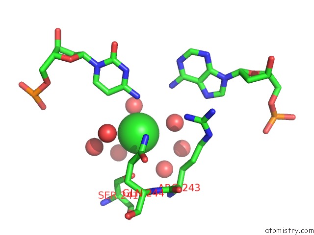
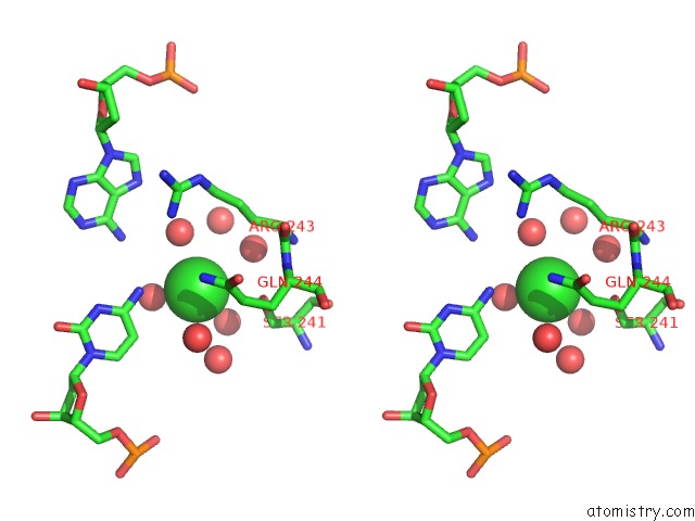
Chlorine binding site 1 out of 3 in 3ubt
Go back to
Chlorine binding site 1 out
of 3 in the Crystal Structure of C71S Mutant of Dna Cytosine-5 Methyltransferase M.Haeiii Bound to Dna
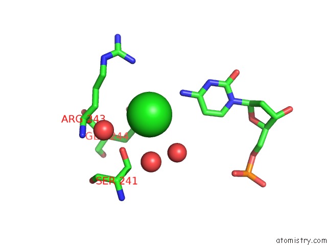
Mono view
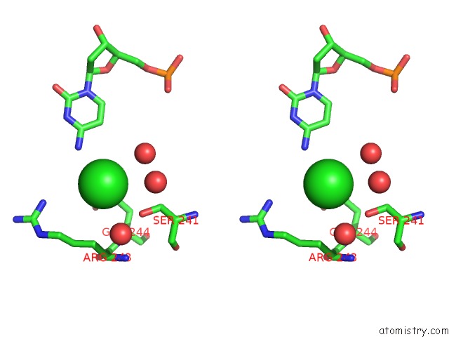
Stereo pair view
Mono view
Stereo pair view
A full contact list of Chlorine with other atoms in the Cl binding
site number 1 of Crystal Structure of C71S Mutant of Dna Cytosine-5 Methyltransferase M.Haeiii Bound to Dna within 5.0Å range:
|
Chlorine binding site 2 out of 3 in 3ubt
Go back to
Chlorine binding site 2 out
of 3 in the Crystal Structure of C71S Mutant of Dna Cytosine-5 Methyltransferase M.Haeiii Bound to Dna
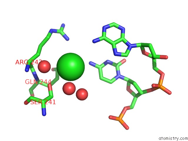
Mono view
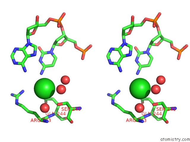
Stereo pair view
Mono view
Stereo pair view
A full contact list of Chlorine with other atoms in the Cl binding
site number 2 of Crystal Structure of C71S Mutant of Dna Cytosine-5 Methyltransferase M.Haeiii Bound to Dna within 5.0Å range:
|
Chlorine binding site 3 out of 3 in 3ubt
Go back to
Chlorine binding site 3 out
of 3 in the Crystal Structure of C71S Mutant of Dna Cytosine-5 Methyltransferase M.Haeiii Bound to Dna
Mono view
Stereo pair view
Mono view
Stereo pair view
A full contact list of Chlorine with other atoms in the Cl binding
site number 3 of Crystal Structure of C71S Mutant of Dna Cytosine-5 Methyltransferase M.Haeiii Bound to Dna within 5.0Å range:
|
Reference:
A.Didovyk,
G.L.Verdine.
Structural Origins of Dna Target Selection and Nucleobase Extrusion By A Dna Cytosine Methyltransferase. J.Biol.Chem. V. 287 40099 2012.
ISSN: ISSN 0021-9258
PubMed: 23012373
DOI: 10.1074/JBC.M112.413054
Page generated: Fri Jul 11 11:11:06 2025
ISSN: ISSN 0021-9258
PubMed: 23012373
DOI: 10.1074/JBC.M112.413054
Last articles
Mg in 1MNDMg in 1MN9
Mg in 1MN7
Mg in 1MMN
Mg in 1MMG
Mg in 1MMD
Mg in 1MJI
Mg in 1MMA
Mg in 1MKJ
Mg in 1MJ5