Chlorine »
PDB 3u6a-3ue5 »
3ue4 »
Chlorine in PDB 3ue4: Structural and Spectroscopic Analysis of the Kinase Inhibitor Bosutinib Binding to the Abl Tyrosine Kinase Domain
Enzymatic activity of Structural and Spectroscopic Analysis of the Kinase Inhibitor Bosutinib Binding to the Abl Tyrosine Kinase Domain
All present enzymatic activity of Structural and Spectroscopic Analysis of the Kinase Inhibitor Bosutinib Binding to the Abl Tyrosine Kinase Domain:
2.7.10.2;
2.7.10.2;
Protein crystallography data
The structure of Structural and Spectroscopic Analysis of the Kinase Inhibitor Bosutinib Binding to the Abl Tyrosine Kinase Domain, PDB code: 3ue4
was solved by
S.G.Boxer,
N.M.Levinson,
with X-Ray Crystallography technique. A brief refinement statistics is given in the table below:
| Resolution Low / High (Å) | 63.82 / 2.42 |
| Space group | P 2 21 21 |
| Cell size a, b, c (Å), α, β, γ (°) | 56.860, 113.760, 127.640, 90.00, 90.00, 90.00 |
| R / Rfree (%) | 18.8 / 24.9 |
Chlorine Binding Sites:
The binding sites of Chlorine atom in the Structural and Spectroscopic Analysis of the Kinase Inhibitor Bosutinib Binding to the Abl Tyrosine Kinase Domain
(pdb code 3ue4). This binding sites where shown within
5.0 Angstroms radius around Chlorine atom.
In total 4 binding sites of Chlorine where determined in the Structural and Spectroscopic Analysis of the Kinase Inhibitor Bosutinib Binding to the Abl Tyrosine Kinase Domain, PDB code: 3ue4:
Jump to Chlorine binding site number: 1; 2; 3; 4;
In total 4 binding sites of Chlorine where determined in the Structural and Spectroscopic Analysis of the Kinase Inhibitor Bosutinib Binding to the Abl Tyrosine Kinase Domain, PDB code: 3ue4:
Jump to Chlorine binding site number: 1; 2; 3; 4;
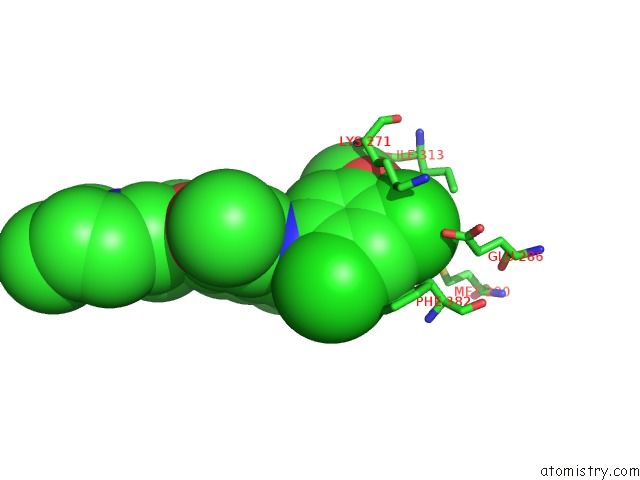
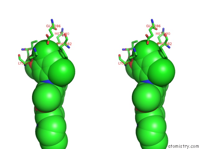
Chlorine binding site 1 out of 4 in 3ue4
Go back to
Chlorine binding site 1 out
of 4 in the Structural and Spectroscopic Analysis of the Kinase Inhibitor Bosutinib Binding to the Abl Tyrosine Kinase Domain
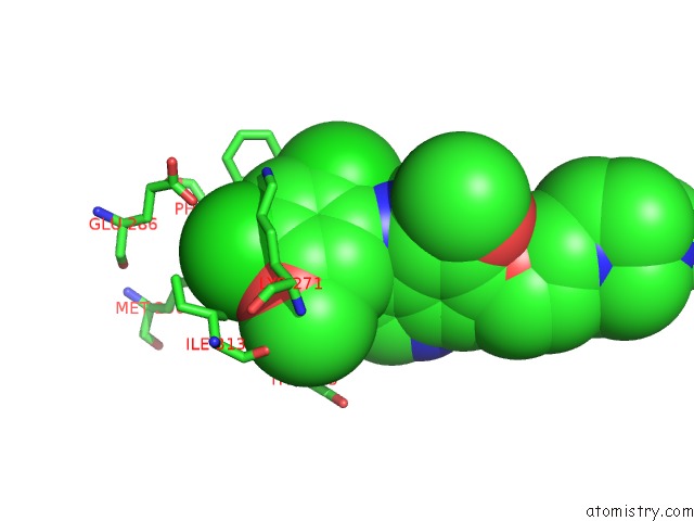
Mono view
Stereo pair view
Mono view
Stereo pair view
A full contact list of Chlorine with other atoms in the Cl binding
site number 1 of Structural and Spectroscopic Analysis of the Kinase Inhibitor Bosutinib Binding to the Abl Tyrosine Kinase Domain within 5.0Å range:
|
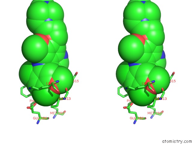
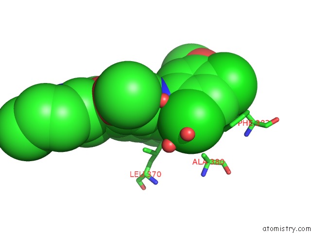
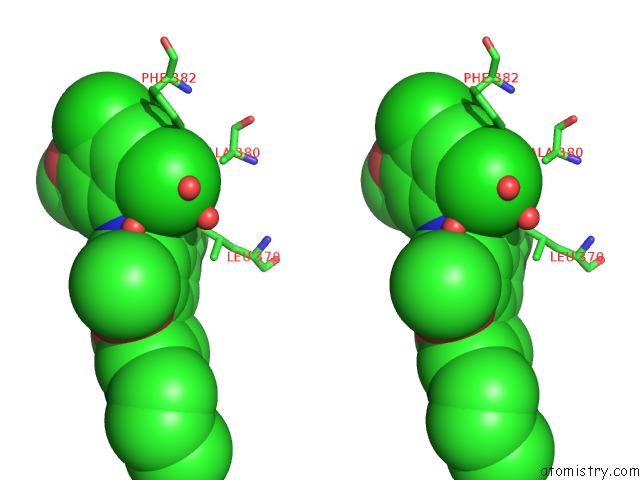
Chlorine binding site 2 out of 4 in 3ue4
Go back to
Chlorine binding site 2 out
of 4 in the Structural and Spectroscopic Analysis of the Kinase Inhibitor Bosutinib Binding to the Abl Tyrosine Kinase Domain
Mono view
Stereo pair view
Mono view
Stereo pair view
A full contact list of Chlorine with other atoms in the Cl binding
site number 2 of Structural and Spectroscopic Analysis of the Kinase Inhibitor Bosutinib Binding to the Abl Tyrosine Kinase Domain within 5.0Å range:
|
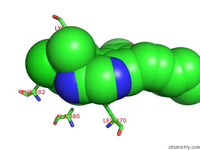
Chlorine binding site 3 out of 4 in 3ue4
Go back to
Chlorine binding site 3 out
of 4 in the Structural and Spectroscopic Analysis of the Kinase Inhibitor Bosutinib Binding to the Abl Tyrosine Kinase Domain
Mono view
Stereo pair view
Mono view
Stereo pair view
A full contact list of Chlorine with other atoms in the Cl binding
site number 3 of Structural and Spectroscopic Analysis of the Kinase Inhibitor Bosutinib Binding to the Abl Tyrosine Kinase Domain within 5.0Å range:
|
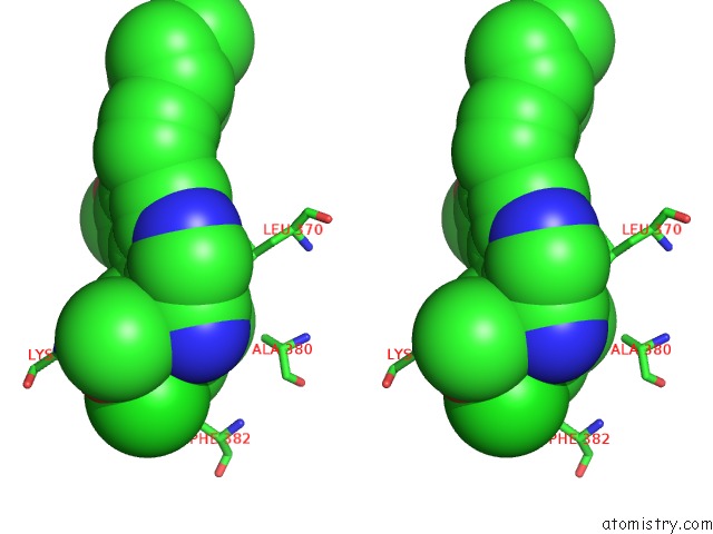
Chlorine binding site 4 out of 4 in 3ue4
Go back to
Chlorine binding site 4 out
of 4 in the Structural and Spectroscopic Analysis of the Kinase Inhibitor Bosutinib Binding to the Abl Tyrosine Kinase Domain
Mono view
Stereo pair view
Mono view
Stereo pair view
A full contact list of Chlorine with other atoms in the Cl binding
site number 4 of Structural and Spectroscopic Analysis of the Kinase Inhibitor Bosutinib Binding to the Abl Tyrosine Kinase Domain within 5.0Å range:
|
Reference:
N.M.Levinson,
S.G.Boxer.
Structural and Spectroscopic Analysis of the Kinase Inhibitor Bosutinib and An Isomer of Bosutinib Binding to the Abl Tyrosine Kinase Domain. Plos One V. 7 29828 2012.
ISSN: ESSN 1932-6203
PubMed: 22493660
DOI: 10.1371/JOURNAL.PONE.0029828
Page generated: Fri Jul 11 11:12:24 2025
ISSN: ESSN 1932-6203
PubMed: 22493660
DOI: 10.1371/JOURNAL.PONE.0029828
Last articles
Mg in 1R0AMg in 1R03
Mg in 1QZR
Mg in 1QU2
Mg in 1QYF
Mg in 1QVJ
Mg in 1QVI
Mg in 1QV9
Mg in 1QSI
Mg in 1QSY