Chlorine »
PDB 3ue8-3unh »
3uhf »
Chlorine in PDB 3uhf: Crystal Structure of Glutamate Racemase From Campylobacter Jejuni Subsp. Jejuni
Enzymatic activity of Crystal Structure of Glutamate Racemase From Campylobacter Jejuni Subsp. Jejuni
All present enzymatic activity of Crystal Structure of Glutamate Racemase From Campylobacter Jejuni Subsp. Jejuni:
5.1.1.3;
5.1.1.3;
Protein crystallography data
The structure of Crystal Structure of Glutamate Racemase From Campylobacter Jejuni Subsp. Jejuni, PDB code: 3uhf
was solved by
N.Maltseva,
R.Mulligan,
K.Kwon,
Y.Kim,
W.F.Anderson,
A.Joachimiak,
Centerfor Structural Genomics Of Infectious Diseases (Csgid),
with X-Ray Crystallography technique. A brief refinement statistics is given in the table below:
| Resolution Low / High (Å) | 44.95 / 1.83 |
| Space group | P 21 21 21 |
| Cell size a, b, c (Å), α, β, γ (°) | 47.453, 88.839, 140.177, 90.00, 90.00, 90.00 |
| R / Rfree (%) | 16.2 / 19 |
Chlorine Binding Sites:
The binding sites of Chlorine atom in the Crystal Structure of Glutamate Racemase From Campylobacter Jejuni Subsp. Jejuni
(pdb code 3uhf). This binding sites where shown within
5.0 Angstroms radius around Chlorine atom.
In total 2 binding sites of Chlorine where determined in the Crystal Structure of Glutamate Racemase From Campylobacter Jejuni Subsp. Jejuni, PDB code: 3uhf:
Jump to Chlorine binding site number: 1; 2;
In total 2 binding sites of Chlorine where determined in the Crystal Structure of Glutamate Racemase From Campylobacter Jejuni Subsp. Jejuni, PDB code: 3uhf:
Jump to Chlorine binding site number: 1; 2;
Chlorine binding site 1 out of 2 in 3uhf
Go back to
Chlorine binding site 1 out
of 2 in the Crystal Structure of Glutamate Racemase From Campylobacter Jejuni Subsp. Jejuni
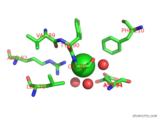
Mono view
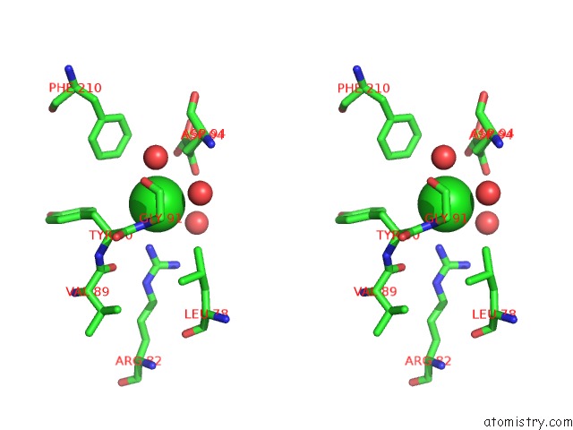
Stereo pair view
Mono view
Stereo pair view
A full contact list of Chlorine with other atoms in the Cl binding
site number 1 of Crystal Structure of Glutamate Racemase From Campylobacter Jejuni Subsp. Jejuni within 5.0Å range:
|
Chlorine binding site 2 out of 2 in 3uhf
Go back to
Chlorine binding site 2 out
of 2 in the Crystal Structure of Glutamate Racemase From Campylobacter Jejuni Subsp. Jejuni
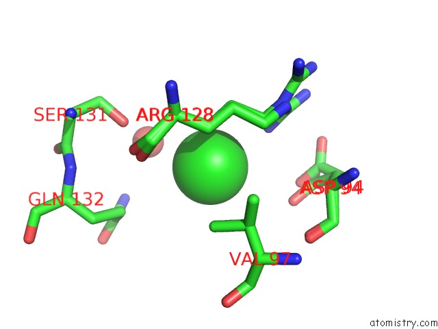
Mono view
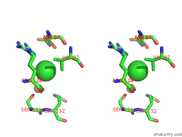
Stereo pair view
Mono view
Stereo pair view
A full contact list of Chlorine with other atoms in the Cl binding
site number 2 of Crystal Structure of Glutamate Racemase From Campylobacter Jejuni Subsp. Jejuni within 5.0Å range:
|
Reference:
N.Maltseva,
R.Mulligan,
K.Kwon,
Y.Kim,
W.F.Anderson,
A.Joachimiak,
Csgid.
Crystal Structure of Glutamate Racemase From Campylobacter Jejuni Subsp. Jejuni To Be Published.
Page generated: Fri Jul 11 11:15:48 2025
Last articles
Mg in 6U9HMg in 6UBR
Mg in 6UBN
Mg in 6UBQ
Mg in 6U9Q
Mg in 6U91
Mg in 6U8Q
Mg in 6U8W
Mg in 6U8X
Mg in 6U8V