Chlorine »
PDB 3ue8-3unh »
3uix »
Chlorine in PDB 3uix: Crystal Structure of PIM1 Kinase in Complex with Small Molecule Inhibitor
Enzymatic activity of Crystal Structure of PIM1 Kinase in Complex with Small Molecule Inhibitor
All present enzymatic activity of Crystal Structure of PIM1 Kinase in Complex with Small Molecule Inhibitor:
2.7.11.1;
2.7.11.1;
Protein crystallography data
The structure of Crystal Structure of PIM1 Kinase in Complex with Small Molecule Inhibitor, PDB code: 3uix
was solved by
L.J.Parker,
with X-Ray Crystallography technique. A brief refinement statistics is given in the table below:
| Resolution Low / High (Å) | 29.14 / 2.20 |
| Space group | P 65 |
| Cell size a, b, c (Å), α, β, γ (°) | 97.006, 97.006, 80.898, 90.00, 90.00, 120.00 |
| R / Rfree (%) | 16.6 / 20.1 |
Other elements in 3uix:
The structure of Crystal Structure of PIM1 Kinase in Complex with Small Molecule Inhibitor also contains other interesting chemical elements:
| Calcium | (Ca) | 1 atom |
Chlorine Binding Sites:
The binding sites of Chlorine atom in the Crystal Structure of PIM1 Kinase in Complex with Small Molecule Inhibitor
(pdb code 3uix). This binding sites where shown within
5.0 Angstroms radius around Chlorine atom.
In total 2 binding sites of Chlorine where determined in the Crystal Structure of PIM1 Kinase in Complex with Small Molecule Inhibitor, PDB code: 3uix:
Jump to Chlorine binding site number: 1; 2;
In total 2 binding sites of Chlorine where determined in the Crystal Structure of PIM1 Kinase in Complex with Small Molecule Inhibitor, PDB code: 3uix:
Jump to Chlorine binding site number: 1; 2;
Chlorine binding site 1 out of 2 in 3uix
Go back to
Chlorine binding site 1 out
of 2 in the Crystal Structure of PIM1 Kinase in Complex with Small Molecule Inhibitor
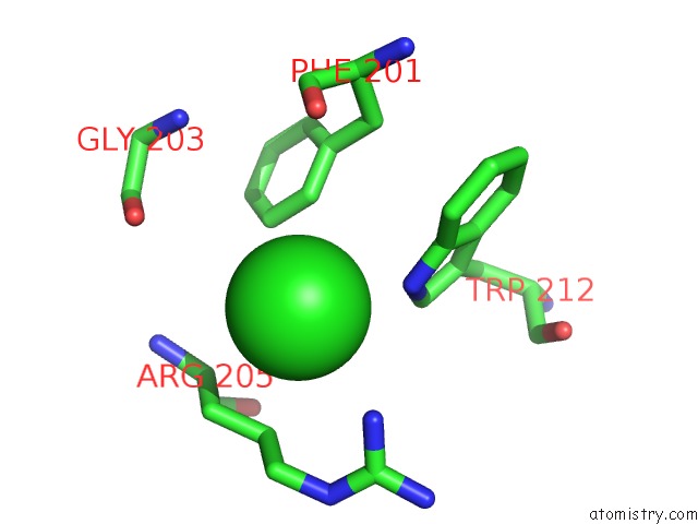
Mono view
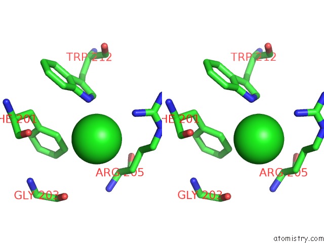
Stereo pair view
Mono view
Stereo pair view
A full contact list of Chlorine with other atoms in the Cl binding
site number 1 of Crystal Structure of PIM1 Kinase in Complex with Small Molecule Inhibitor within 5.0Å range:
|
Chlorine binding site 2 out of 2 in 3uix
Go back to
Chlorine binding site 2 out
of 2 in the Crystal Structure of PIM1 Kinase in Complex with Small Molecule Inhibitor
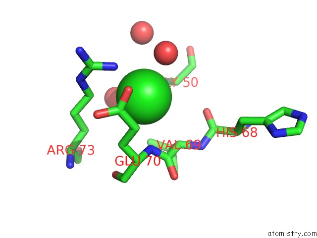
Mono view
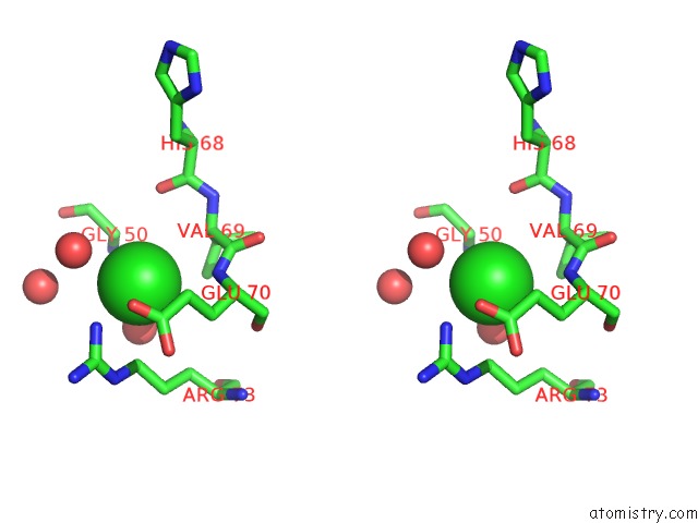
Stereo pair view
Mono view
Stereo pair view
A full contact list of Chlorine with other atoms in the Cl binding
site number 2 of Crystal Structure of PIM1 Kinase in Complex with Small Molecule Inhibitor within 5.0Å range:
|
Reference:
K.Tsuganezawa,
H.Watanabe,
L.Parker,
H.Yuki,
S.Taruya,
Y.Nakagawa,
D.Kamei,
M.Mori,
N.Ogawa,
Y.Tomabechi,
N.Handa,
T.Honma,
S.Yokoyama,
H.Kojima,
T.Okabe,
T.Nagano,
A.Tanaka.
A Novel Pim-1 Kinase Inhibitor Targeting Residues That Bind the Substrate Peptide. J.Mol.Biol. V. 417 240 2012.
ISSN: ISSN 0022-2836
PubMed: 22306408
DOI: 10.1016/J.JMB.2012.01.036
Page generated: Fri Jul 11 11:16:31 2025
ISSN: ISSN 0022-2836
PubMed: 22306408
DOI: 10.1016/J.JMB.2012.01.036
Last articles
Mg in 6C3OMg in 6C2C
Mg in 6C2X
Mg in 6C25
Mg in 6C1X
Mg in 6C1H
Mg in 6C1G
Mg in 6C1D
Mg in 6C1J
Mg in 6C0V