Chlorine »
PDB 3unk-3uwd »
3uss »
Chlorine in PDB 3uss: Crystal Structure of Cysteine Dioxygenase From Pseudomonas Aeruginosa
Enzymatic activity of Crystal Structure of Cysteine Dioxygenase From Pseudomonas Aeruginosa
All present enzymatic activity of Crystal Structure of Cysteine Dioxygenase From Pseudomonas Aeruginosa:
1.13.11.20;
1.13.11.20;
Protein crystallography data
The structure of Crystal Structure of Cysteine Dioxygenase From Pseudomonas Aeruginosa, PDB code: 3uss
was solved by
S.M.Wilbanks,
E.P.Tchesnokov,
G.N.L.Jameson,
with X-Ray Crystallography technique. A brief refinement statistics is given in the table below:
| Resolution Low / High (Å) | 53.30 / 2.70 |
| Space group | P 21 21 21 |
| Cell size a, b, c (Å), α, β, γ (°) | 53.330, 86.790, 123.000, 90.00, 90.00, 90.00 |
| R / Rfree (%) | 20.6 / 27.7 |
Other elements in 3uss:
The structure of Crystal Structure of Cysteine Dioxygenase From Pseudomonas Aeruginosa also contains other interesting chemical elements:
| Iron | (Fe) | 2 atoms |
| Sodium | (Na) | 4 atoms |
Chlorine Binding Sites:
The binding sites of Chlorine atom in the Crystal Structure of Cysteine Dioxygenase From Pseudomonas Aeruginosa
(pdb code 3uss). This binding sites where shown within
5.0 Angstroms radius around Chlorine atom.
In total only one binding site of Chlorine was determined in the Crystal Structure of Cysteine Dioxygenase From Pseudomonas Aeruginosa, PDB code: 3uss:
In total only one binding site of Chlorine was determined in the Crystal Structure of Cysteine Dioxygenase From Pseudomonas Aeruginosa, PDB code: 3uss:
Chlorine binding site 1 out of 1 in 3uss
Go back to
Chlorine binding site 1 out
of 1 in the Crystal Structure of Cysteine Dioxygenase From Pseudomonas Aeruginosa
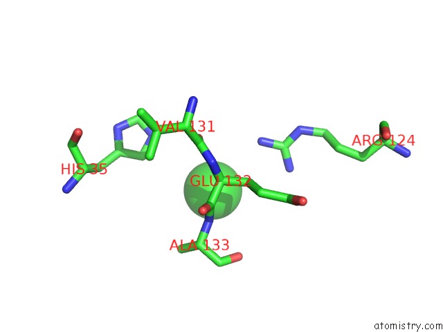
Mono view
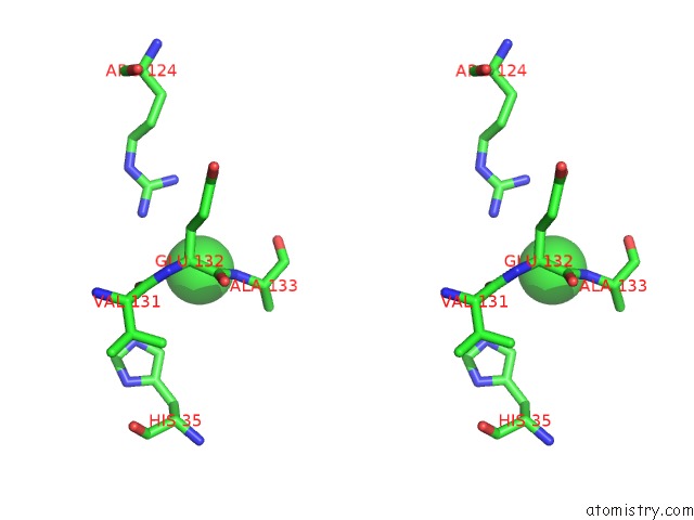
Stereo pair view
Mono view
Stereo pair view
A full contact list of Chlorine with other atoms in the Cl binding
site number 1 of Crystal Structure of Cysteine Dioxygenase From Pseudomonas Aeruginosa within 5.0Å range:
|
Reference:
G.N.L.Jameson,
E.P.Tchesnokov,
E.Siakkou,
S.M.Wilbanks.
Crystal Structure, Kinetics and M Ssbauer Spectroscopic Studies of A Bacterial Cysteine Dioxygenase To Be Published.
Page generated: Fri Jul 11 11:27:33 2025
Last articles
Mg in 2UUBMg in 2UUC
Mg in 2UX5
Mg in 2UX4
Mg in 2UX3
Mg in 2UWW
Mg in 2UWV
Mg in 2UWU
Mg in 2UWT
Mg in 2UUA