Chlorine »
PDB 3vm6-3vwd »
3voc »
Chlorine in PDB 3voc: Crystal Structure of the Catalytic Domain of Beta-Amylase From Paenibacillus Polymyxa
Enzymatic activity of Crystal Structure of the Catalytic Domain of Beta-Amylase From Paenibacillus Polymyxa
All present enzymatic activity of Crystal Structure of the Catalytic Domain of Beta-Amylase From Paenibacillus Polymyxa:
3.2.1.2;
3.2.1.2;
Protein crystallography data
The structure of Crystal Structure of the Catalytic Domain of Beta-Amylase From Paenibacillus Polymyxa, PDB code: 3voc
was solved by
S.Nishimura,
T.Fujioka,
T.Nakaniwa,
T.Tada,
with X-Ray Crystallography technique. A brief refinement statistics is given in the table below:
| Resolution Low / High (Å) | 41.00 / 1.95 |
| Space group | P 21 21 21 |
| Cell size a, b, c (Å), α, β, γ (°) | 61.028, 68.519, 93.904, 90.00, 90.00, 90.00 |
| R / Rfree (%) | 15.3 / 19 |
Other elements in 3voc:
The structure of Crystal Structure of the Catalytic Domain of Beta-Amylase From Paenibacillus Polymyxa also contains other interesting chemical elements:
| Calcium | (Ca) | 1 atom |
Chlorine Binding Sites:
The binding sites of Chlorine atom in the Crystal Structure of the Catalytic Domain of Beta-Amylase From Paenibacillus Polymyxa
(pdb code 3voc). This binding sites where shown within
5.0 Angstroms radius around Chlorine atom.
In total only one binding site of Chlorine was determined in the Crystal Structure of the Catalytic Domain of Beta-Amylase From Paenibacillus Polymyxa, PDB code: 3voc:
In total only one binding site of Chlorine was determined in the Crystal Structure of the Catalytic Domain of Beta-Amylase From Paenibacillus Polymyxa, PDB code: 3voc:
Chlorine binding site 1 out of 1 in 3voc
Go back to
Chlorine binding site 1 out
of 1 in the Crystal Structure of the Catalytic Domain of Beta-Amylase From Paenibacillus Polymyxa
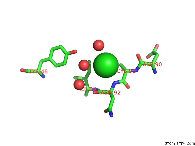
Mono view
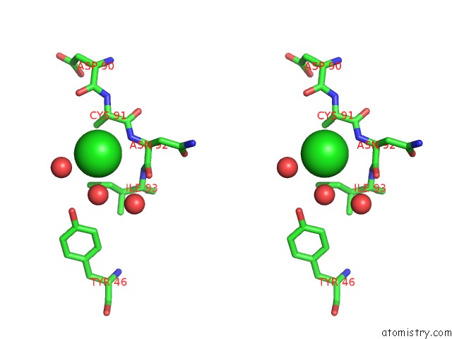
Stereo pair view
Mono view
Stereo pair view
A full contact list of Chlorine with other atoms in the Cl binding
site number 1 of Crystal Structure of the Catalytic Domain of Beta-Amylase From Paenibacillus Polymyxa within 5.0Å range:
|
Reference:
S.Nishimura,
T.Fujioka,
R.Takahashi,
T.Nakaniwa,
H.Fukada,
T.Inui,
T.Tada,
T.Kawaguchi,
J.Sumitani.
Structural Analysis By X-Ray Crystallography and Small-Angle Scattering of the Multi-Domain Beta-Amylase From Paenibacillus Polymyxa To Be Published.
Page generated: Fri Jul 11 11:46:51 2025
Last articles
Mg in 9AZLMg in 9AZ6
Mg in 9AZ5
Mg in 9AZ4
Mg in 9AYO
Mg in 9AYP
Mg in 9AW3
Mg in 9AYN
Mg in 9AYI
Mg in 9AXL