Chlorine »
PDB 3zhc-3zoq »
3zoq »
Chlorine in PDB 3zoq: Structure of Bsudg-P56 Complex
Enzymatic activity of Structure of Bsudg-P56 Complex
All present enzymatic activity of Structure of Bsudg-P56 Complex:
3.2.2.27;
3.2.2.27;
Protein crystallography data
The structure of Structure of Bsudg-P56 Complex, PDB code: 3zoq
was solved by
J.I.Banos-Sanz,
L.Mojardin,
J.Sanz-Aparicio,
B.Gonzalez,
M.Salas,
with X-Ray Crystallography technique. A brief refinement statistics is given in the table below:
| Resolution Low / High (Å) | 55.62 / 1.45 |
| Space group | P 21 21 21 |
| Cell size a, b, c (Å), α, β, γ (°) | 53.770, 66.270, 102.280, 90.00, 90.00, 90.00 |
| R / Rfree (%) | 13.9 / 18.2 |
Chlorine Binding Sites:
The binding sites of Chlorine atom in the Structure of Bsudg-P56 Complex
(pdb code 3zoq). This binding sites where shown within
5.0 Angstroms radius around Chlorine atom.
In total only one binding site of Chlorine was determined in the Structure of Bsudg-P56 Complex, PDB code: 3zoq:
In total only one binding site of Chlorine was determined in the Structure of Bsudg-P56 Complex, PDB code: 3zoq:
Chlorine binding site 1 out of 1 in 3zoq
Go back to
Chlorine binding site 1 out
of 1 in the Structure of Bsudg-P56 Complex
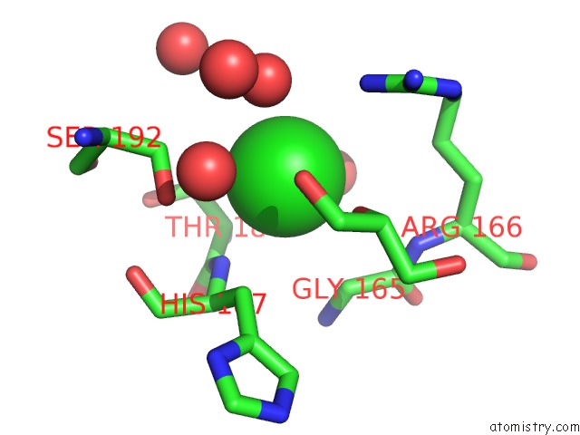
Mono view
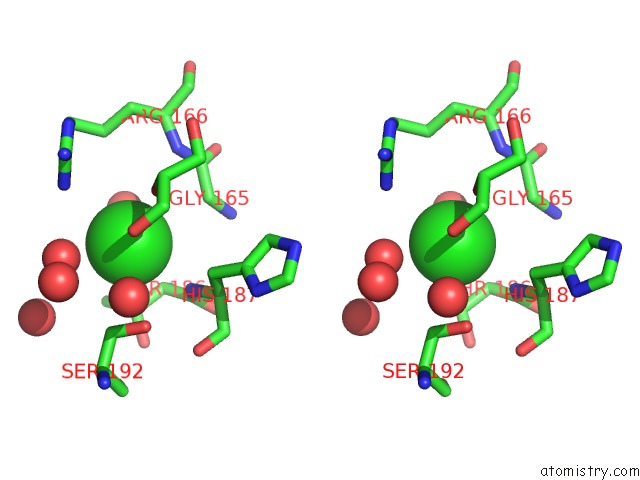
Stereo pair view
Mono view
Stereo pair view
A full contact list of Chlorine with other atoms in the Cl binding
site number 1 of Structure of Bsudg-P56 Complex within 5.0Å range:
|
Reference:
J.I.Banos-Sanz,
L.Mojardin,
J.Sanz-Aparicio,
J.M.Lazaro,
L.Villar,
G.Serrano-Heras,
B.Gonzalez,
M.Salas.
Crystal Structure and Functional Insights Into Uracil-Dna Glycosylase Inhibition By Phage PHI29 Dna Mimic Protein P56 Nucleic Acids Res. V. 41 6761 2013.
ISSN: ISSN 0305-1048
PubMed: 23671337
DOI: 10.1093/NAR/GKT395
Page generated: Fri Jul 11 12:24:15 2025
ISSN: ISSN 0305-1048
PubMed: 23671337
DOI: 10.1093/NAR/GKT395
Last articles
Mg in 6D8AMg in 6D88
Mg in 6D84
Mg in 6D3Q
Mg in 6D83
Mg in 6D71
Mg in 6D5X
Mg in 6D6R
Mg in 6D6Q
Mg in 6D5W