Chlorine »
PDB 4aqi-4azo »
4axw »
Chlorine in PDB 4axw: Crystal Structure of Mouse Cadherin-23 EC1-2 and Protocadherin-15 EC1-2, Form I 2.2A.
Protein crystallography data
The structure of Crystal Structure of Mouse Cadherin-23 EC1-2 and Protocadherin-15 EC1-2, Form I 2.2A., PDB code: 4axw
was solved by
M.Sotomayor,
W.Weihofen,
R.Gaudet,
D.P.Corey,
with X-Ray Crystallography technique. A brief refinement statistics is given in the table below:
| Resolution Low / High (Å) | 41.08 / 2.23 |
| Space group | C 1 2 1 |
| Cell size a, b, c (Å), α, β, γ (°) | 173.649, 40.468, 85.187, 90.00, 102.92, 90.00 |
| R / Rfree (%) | 17.459 / 23.634 |
Other elements in 4axw:
The structure of Crystal Structure of Mouse Cadherin-23 EC1-2 and Protocadherin-15 EC1-2, Form I 2.2A. also contains other interesting chemical elements:
| Potassium | (K) | 1 atom |
| Calcium | (Ca) | 7 atoms |
Chlorine Binding Sites:
The binding sites of Chlorine atom in the Crystal Structure of Mouse Cadherin-23 EC1-2 and Protocadherin-15 EC1-2, Form I 2.2A.
(pdb code 4axw). This binding sites where shown within
5.0 Angstroms radius around Chlorine atom.
In total only one binding site of Chlorine was determined in the Crystal Structure of Mouse Cadherin-23 EC1-2 and Protocadherin-15 EC1-2, Form I 2.2A., PDB code: 4axw:
In total only one binding site of Chlorine was determined in the Crystal Structure of Mouse Cadherin-23 EC1-2 and Protocadherin-15 EC1-2, Form I 2.2A., PDB code: 4axw:
Chlorine binding site 1 out of 1 in 4axw
Go back to
Chlorine binding site 1 out
of 1 in the Crystal Structure of Mouse Cadherin-23 EC1-2 and Protocadherin-15 EC1-2, Form I 2.2A.
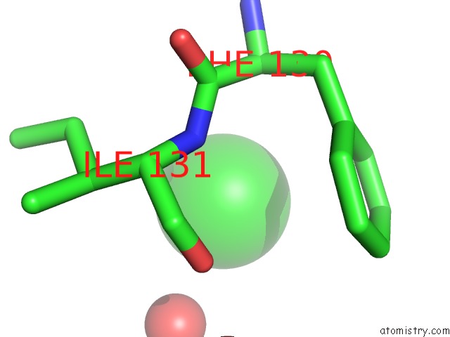
Mono view
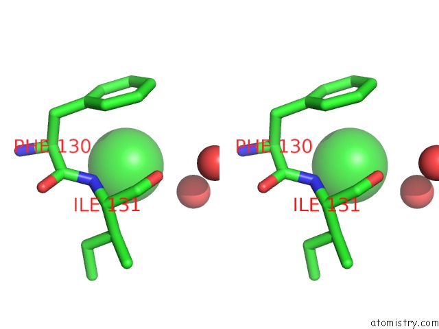
Stereo pair view
Mono view
Stereo pair view
A full contact list of Chlorine with other atoms in the Cl binding
site number 1 of Crystal Structure of Mouse Cadherin-23 EC1-2 and Protocadherin-15 EC1-2, Form I 2.2A. within 5.0Å range:
|
Reference:
M.Sotomayor,
W.Weihofen,
R.Gaudet,
D.P.Corey.
Structure of A Force-Conveying Cadherin Bond Essential For Inner-Ear Mechanotransduction Nature V. 492 128 2012.
ISSN: ISSN 0028-0836
PubMed: 23135401
DOI: 10.1038/NATURE11590
Page generated: Fri Jul 11 13:03:56 2025
ISSN: ISSN 0028-0836
PubMed: 23135401
DOI: 10.1038/NATURE11590
Last articles
Mg in 1YIOMg in 1YHM
Mg in 1YID
Mg in 1YI1
Mg in 1YI0
Mg in 1YHZ
Mg in 1YHL
Mg in 1YHY
Mg in 1YFR
Mg in 1YHN