Chlorine »
PDB 4da5-4dhn »
4dfr »
Chlorine in PDB 4dfr: Crystal Structures of Escherichia Coli and Lactobacillus Casei Dihydrofolate Reductase Refined at 1.7 Angstroms Resolution. I. General Features and Binding of Methotrexate
Enzymatic activity of Crystal Structures of Escherichia Coli and Lactobacillus Casei Dihydrofolate Reductase Refined at 1.7 Angstroms Resolution. I. General Features and Binding of Methotrexate
All present enzymatic activity of Crystal Structures of Escherichia Coli and Lactobacillus Casei Dihydrofolate Reductase Refined at 1.7 Angstroms Resolution. I. General Features and Binding of Methotrexate:
1.5.1.3;
1.5.1.3;
Protein crystallography data
The structure of Crystal Structures of Escherichia Coli and Lactobacillus Casei Dihydrofolate Reductase Refined at 1.7 Angstroms Resolution. I. General Features and Binding of Methotrexate, PDB code: 4dfr
was solved by
D.J.Filman,
D.A.Matthews,
J.T.Bolin,
J.Kraut,
with X-Ray Crystallography technique. A brief refinement statistics is given in the table below:
| Resolution Low / High (Å) | N/A / 1.70 |
| Space group | P 61 |
| Cell size a, b, c (Å), α, β, γ (°) | 93.220, 93.220, 73.560, 90.00, 90.00, 120.00 |
| R / Rfree (%) | 15.5 / n/a |
Other elements in 4dfr:
The structure of Crystal Structures of Escherichia Coli and Lactobacillus Casei Dihydrofolate Reductase Refined at 1.7 Angstroms Resolution. I. General Features and Binding of Methotrexate also contains other interesting chemical elements:
| Calcium | (Ca) | 1 atom |
Chlorine Binding Sites:
The binding sites of Chlorine atom in the Crystal Structures of Escherichia Coli and Lactobacillus Casei Dihydrofolate Reductase Refined at 1.7 Angstroms Resolution. I. General Features and Binding of Methotrexate
(pdb code 4dfr). This binding sites where shown within
5.0 Angstroms radius around Chlorine atom.
In total 2 binding sites of Chlorine where determined in the Crystal Structures of Escherichia Coli and Lactobacillus Casei Dihydrofolate Reductase Refined at 1.7 Angstroms Resolution. I. General Features and Binding of Methotrexate, PDB code: 4dfr:
Jump to Chlorine binding site number: 1; 2;
In total 2 binding sites of Chlorine where determined in the Crystal Structures of Escherichia Coli and Lactobacillus Casei Dihydrofolate Reductase Refined at 1.7 Angstroms Resolution. I. General Features and Binding of Methotrexate, PDB code: 4dfr:
Jump to Chlorine binding site number: 1; 2;
Chlorine binding site 1 out of 2 in 4dfr
Go back to
Chlorine binding site 1 out
of 2 in the Crystal Structures of Escherichia Coli and Lactobacillus Casei Dihydrofolate Reductase Refined at 1.7 Angstroms Resolution. I. General Features and Binding of Methotrexate
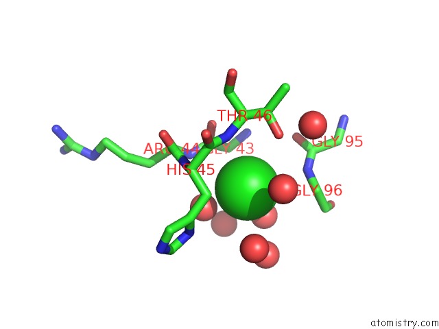
Mono view
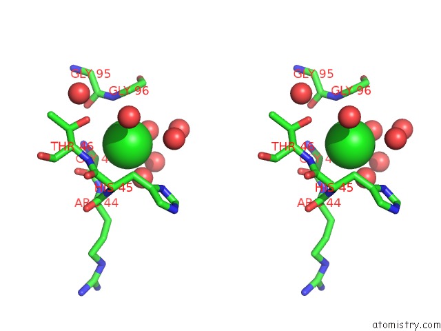
Stereo pair view
Mono view
Stereo pair view
A full contact list of Chlorine with other atoms in the Cl binding
site number 1 of Crystal Structures of Escherichia Coli and Lactobacillus Casei Dihydrofolate Reductase Refined at 1.7 Angstroms Resolution. I. General Features and Binding of Methotrexate within 5.0Å range:
|
Chlorine binding site 2 out of 2 in 4dfr
Go back to
Chlorine binding site 2 out
of 2 in the Crystal Structures of Escherichia Coli and Lactobacillus Casei Dihydrofolate Reductase Refined at 1.7 Angstroms Resolution. I. General Features and Binding of Methotrexate
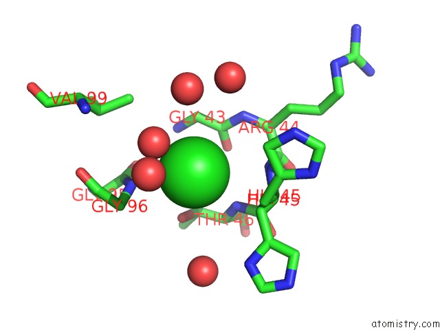
Mono view
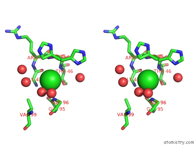
Stereo pair view
Mono view
Stereo pair view
A full contact list of Chlorine with other atoms in the Cl binding
site number 2 of Crystal Structures of Escherichia Coli and Lactobacillus Casei Dihydrofolate Reductase Refined at 1.7 Angstroms Resolution. I. General Features and Binding of Methotrexate within 5.0Å range:
|
Reference:
J.T.Bolin,
D.J.Filman,
D.A.Matthews,
R.C.Hamlin,
J.Kraut.
Crystal Structures of Escherichia Coli and Lactobacillus Casei Dihydrofolate Reductase Refined at 1.7 A Resolution. I. General Features and Binding of Methotrexate. J.Biol.Chem. V. 257 13650 1982.
ISSN: ISSN 0021-9258
PubMed: 6815178
Page generated: Fri Jul 11 14:21:27 2025
ISSN: ISSN 0021-9258
PubMed: 6815178
Last articles
Mg in 4NXIMg in 4NX8
Mg in 4NV0
Mg in 4NWI
Mg in 4NX5
Mg in 4NV3
Mg in 4NW7
Mg in 4NRU
Mg in 4NST
Mg in 4NUA