Chlorine »
PDB 4dq9-4e02 »
4dxv »
Chlorine in PDB 4dxv: Crystal Structure of Dihydrodipicolinate Synthase From Acinetobacter Baumannii Complexed with Mg and Cl Ions at 1.80 A Resolution
Enzymatic activity of Crystal Structure of Dihydrodipicolinate Synthase From Acinetobacter Baumannii Complexed with Mg and Cl Ions at 1.80 A Resolution
All present enzymatic activity of Crystal Structure of Dihydrodipicolinate Synthase From Acinetobacter Baumannii Complexed with Mg and Cl Ions at 1.80 A Resolution:
4.2.1.52;
4.2.1.52;
Protein crystallography data
The structure of Crystal Structure of Dihydrodipicolinate Synthase From Acinetobacter Baumannii Complexed with Mg and Cl Ions at 1.80 A Resolution, PDB code: 4dxv
was solved by
M.Kumar,
S.Kaushik,
M.Sinha,
P.Kaur,
S.Sharma,
T.P.Singh,
with X-Ray Crystallography technique. A brief refinement statistics is given in the table below:
| Resolution Low / High (Å) | 61.24 / 1.80 |
| Space group | P 1 21 1 |
| Cell size a, b, c (Å), α, β, γ (°) | 51.350, 122.470, 52.330, 90.00, 115.92, 90.00 |
| R / Rfree (%) | 15.2 / 19.9 |
Other elements in 4dxv:
The structure of Crystal Structure of Dihydrodipicolinate Synthase From Acinetobacter Baumannii Complexed with Mg and Cl Ions at 1.80 A Resolution also contains other interesting chemical elements:
| Magnesium | (Mg) | 1 atom |
Chlorine Binding Sites:
The binding sites of Chlorine atom in the Crystal Structure of Dihydrodipicolinate Synthase From Acinetobacter Baumannii Complexed with Mg and Cl Ions at 1.80 A Resolution
(pdb code 4dxv). This binding sites where shown within
5.0 Angstroms radius around Chlorine atom.
In total 2 binding sites of Chlorine where determined in the Crystal Structure of Dihydrodipicolinate Synthase From Acinetobacter Baumannii Complexed with Mg and Cl Ions at 1.80 A Resolution, PDB code: 4dxv:
Jump to Chlorine binding site number: 1; 2;
In total 2 binding sites of Chlorine where determined in the Crystal Structure of Dihydrodipicolinate Synthase From Acinetobacter Baumannii Complexed with Mg and Cl Ions at 1.80 A Resolution, PDB code: 4dxv:
Jump to Chlorine binding site number: 1; 2;
Chlorine binding site 1 out of 2 in 4dxv
Go back to
Chlorine binding site 1 out
of 2 in the Crystal Structure of Dihydrodipicolinate Synthase From Acinetobacter Baumannii Complexed with Mg and Cl Ions at 1.80 A Resolution
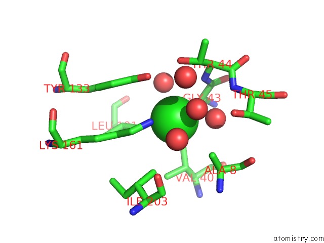
Mono view
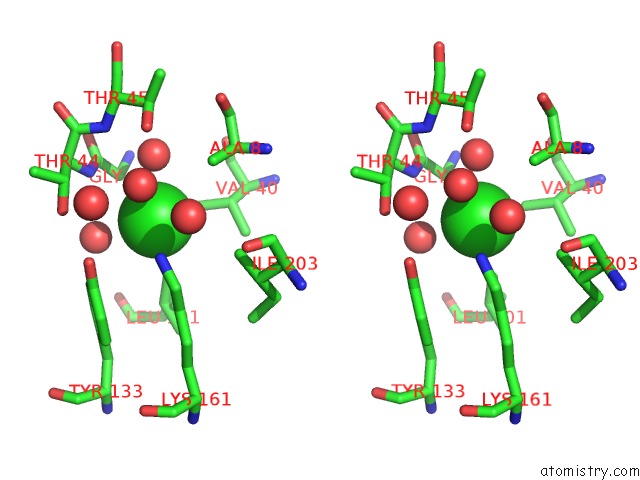
Stereo pair view
Mono view
Stereo pair view
A full contact list of Chlorine with other atoms in the Cl binding
site number 1 of Crystal Structure of Dihydrodipicolinate Synthase From Acinetobacter Baumannii Complexed with Mg and Cl Ions at 1.80 A Resolution within 5.0Å range:
|
Chlorine binding site 2 out of 2 in 4dxv
Go back to
Chlorine binding site 2 out
of 2 in the Crystal Structure of Dihydrodipicolinate Synthase From Acinetobacter Baumannii Complexed with Mg and Cl Ions at 1.80 A Resolution
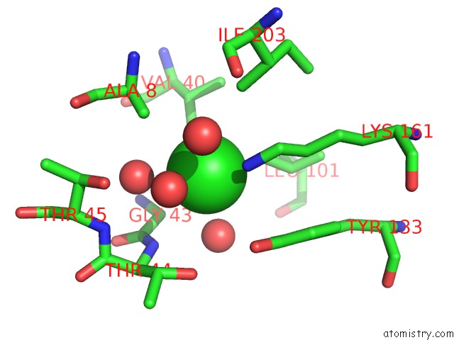
Mono view
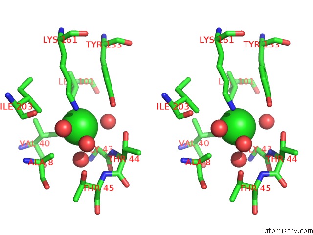
Stereo pair view
Mono view
Stereo pair view
A full contact list of Chlorine with other atoms in the Cl binding
site number 2 of Crystal Structure of Dihydrodipicolinate Synthase From Acinetobacter Baumannii Complexed with Mg and Cl Ions at 1.80 A Resolution within 5.0Å range:
|
Reference:
M.Kumar,
S.Kaushik,
M.Sinha,
P.Kaur,
S.Sharma,
T.P.Singh.
Crystal Structure of Dihydrodipicolinate Synthase From Acinetobacter Baumannii Complexed with Mg and Cl Ions at 1.80 A Resolution To Be Published.
Page generated: Fri Jul 11 14:33:08 2025
Last articles
Mg in 5BYAMg in 5BXN
Mg in 5BYB
Mg in 5BXL
Mg in 5BY0
Mg in 5BTP
Mg in 5BXY
Mg in 5BXW
Mg in 5BXQ
Mg in 5BTN