Chlorine »
PDB 4eu6-4f1a »
4exl »
Chlorine in PDB 4exl: Crystal Structure of Phosphate Abc Transporter, Periplasmic Phosphate- Binding Protein Psts 1 (PBP1) From Streptococcus Pneumoniae Canada MDR_19A
Protein crystallography data
The structure of Crystal Structure of Phosphate Abc Transporter, Periplasmic Phosphate- Binding Protein Psts 1 (PBP1) From Streptococcus Pneumoniae Canada MDR_19A, PDB code: 4exl
was solved by
P.J.Stogios,
Z.Wawrzak,
M.Kudritska,
G.Minasov,
V.Yim,
A.Savchenko,
W.F.Anderson,
Center For Structural Genomics Of Infectious Diseases(Csgid),
with X-Ray Crystallography technique. A brief refinement statistics is given in the table below:
| Resolution Low / High (Å) | 24.85 / 1.70 |
| Space group | P 1 21 1 |
| Cell size a, b, c (Å), α, β, γ (°) | 56.633, 76.501, 111.066, 90.00, 93.88, 90.00 |
| R / Rfree (%) | 16.5 / 21.1 |
Other elements in 4exl:
The structure of Crystal Structure of Phosphate Abc Transporter, Periplasmic Phosphate- Binding Protein Psts 1 (PBP1) From Streptococcus Pneumoniae Canada MDR_19A also contains other interesting chemical elements:
| Magnesium | (Mg) | 6 atoms |
Chlorine Binding Sites:
The binding sites of Chlorine atom in the Crystal Structure of Phosphate Abc Transporter, Periplasmic Phosphate- Binding Protein Psts 1 (PBP1) From Streptococcus Pneumoniae Canada MDR_19A
(pdb code 4exl). This binding sites where shown within
5.0 Angstroms radius around Chlorine atom.
In total 7 binding sites of Chlorine where determined in the Crystal Structure of Phosphate Abc Transporter, Periplasmic Phosphate- Binding Protein Psts 1 (PBP1) From Streptococcus Pneumoniae Canada MDR_19A, PDB code: 4exl:
Jump to Chlorine binding site number: 1; 2; 3; 4; 5; 6; 7;
In total 7 binding sites of Chlorine where determined in the Crystal Structure of Phosphate Abc Transporter, Periplasmic Phosphate- Binding Protein Psts 1 (PBP1) From Streptococcus Pneumoniae Canada MDR_19A, PDB code: 4exl:
Jump to Chlorine binding site number: 1; 2; 3; 4; 5; 6; 7;
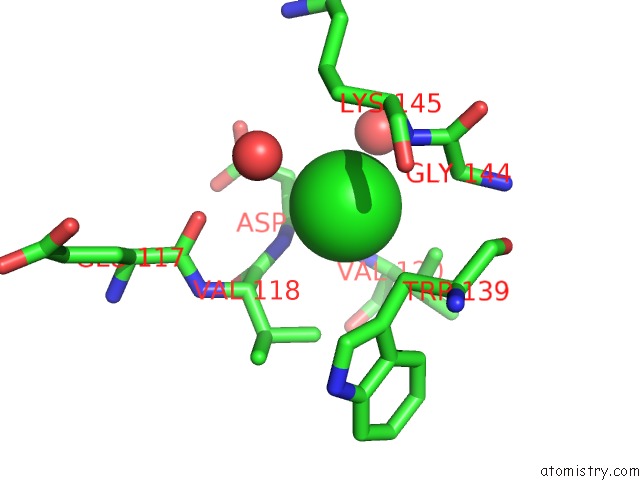
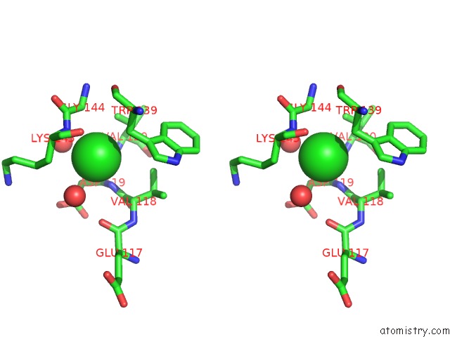
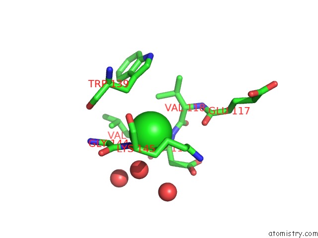
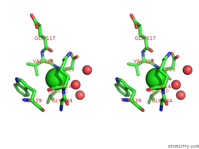
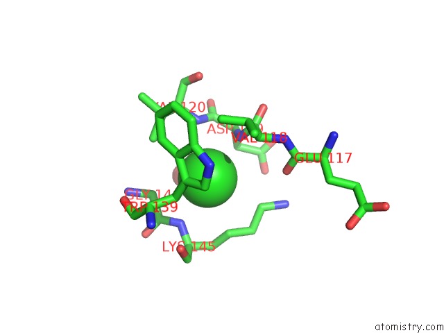
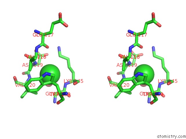
Chlorine binding site 1 out of 7 in 4exl
Go back to
Chlorine binding site 1 out
of 7 in the Crystal Structure of Phosphate Abc Transporter, Periplasmic Phosphate- Binding Protein Psts 1 (PBP1) From Streptococcus Pneumoniae Canada MDR_19A
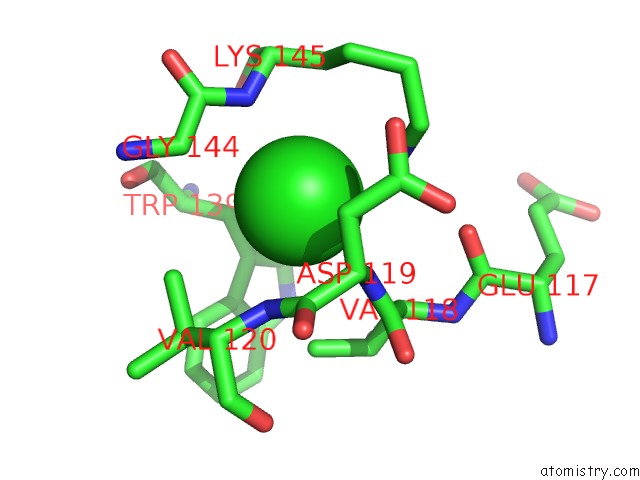
Mono view
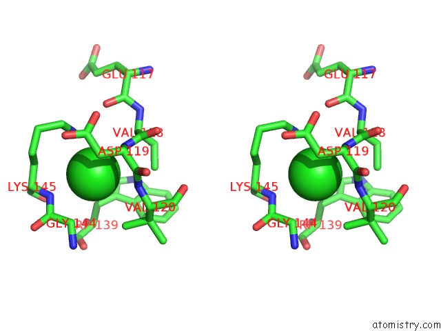
Stereo pair view
Mono view
Stereo pair view
A full contact list of Chlorine with other atoms in the Cl binding
site number 1 of Crystal Structure of Phosphate Abc Transporter, Periplasmic Phosphate- Binding Protein Psts 1 (PBP1) From Streptococcus Pneumoniae Canada MDR_19A within 5.0Å range:
|
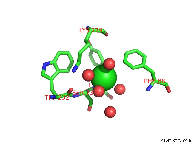
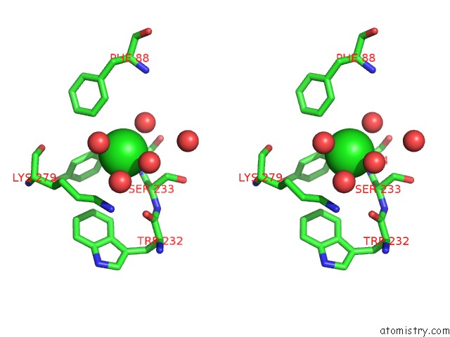
Chlorine binding site 2 out of 7 in 4exl
Go back to
Chlorine binding site 2 out
of 7 in the Crystal Structure of Phosphate Abc Transporter, Periplasmic Phosphate- Binding Protein Psts 1 (PBP1) From Streptococcus Pneumoniae Canada MDR_19A
Mono view
Stereo pair view
Mono view
Stereo pair view
A full contact list of Chlorine with other atoms in the Cl binding
site number 2 of Crystal Structure of Phosphate Abc Transporter, Periplasmic Phosphate- Binding Protein Psts 1 (PBP1) From Streptococcus Pneumoniae Canada MDR_19A within 5.0Å range:
|
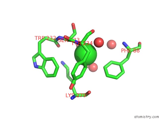
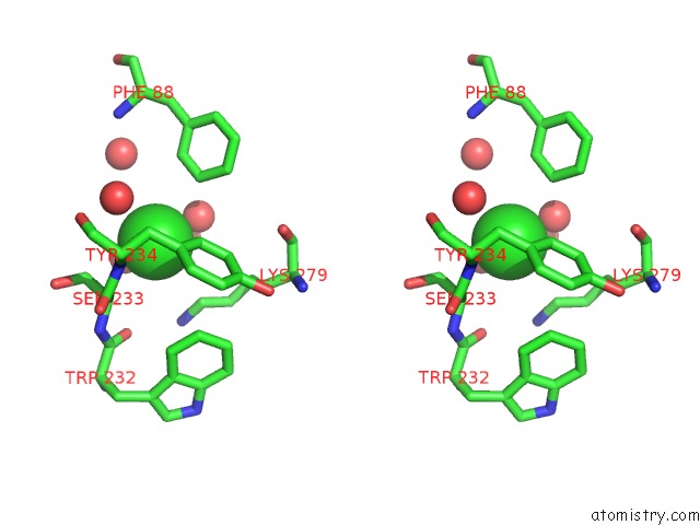
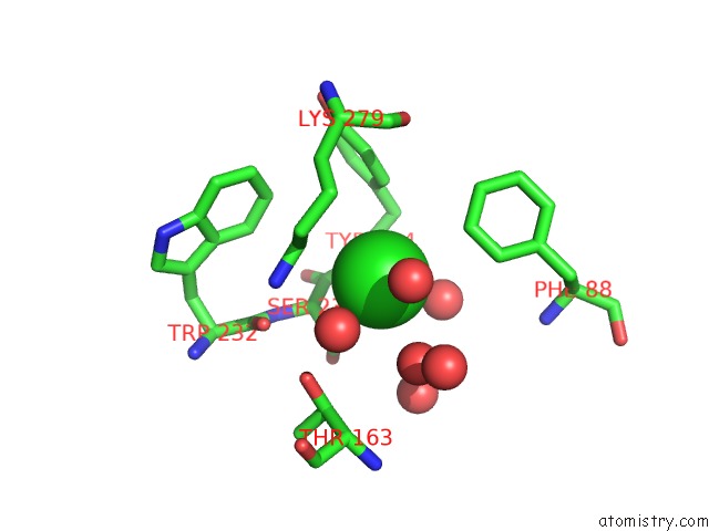
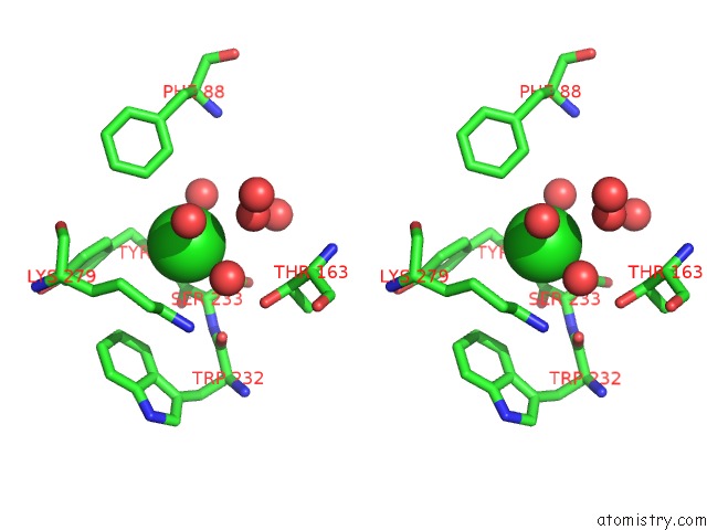
Chlorine binding site 3 out of 7 in 4exl
Go back to
Chlorine binding site 3 out
of 7 in the Crystal Structure of Phosphate Abc Transporter, Periplasmic Phosphate- Binding Protein Psts 1 (PBP1) From Streptococcus Pneumoniae Canada MDR_19A
Mono view
Stereo pair view
Mono view
Stereo pair view
A full contact list of Chlorine with other atoms in the Cl binding
site number 3 of Crystal Structure of Phosphate Abc Transporter, Periplasmic Phosphate- Binding Protein Psts 1 (PBP1) From Streptococcus Pneumoniae Canada MDR_19A within 5.0Å range:
|
Chlorine binding site 4 out of 7 in 4exl
Go back to
Chlorine binding site 4 out
of 7 in the Crystal Structure of Phosphate Abc Transporter, Periplasmic Phosphate- Binding Protein Psts 1 (PBP1) From Streptococcus Pneumoniae Canada MDR_19A
Mono view
Stereo pair view
Mono view
Stereo pair view
A full contact list of Chlorine with other atoms in the Cl binding
site number 4 of Crystal Structure of Phosphate Abc Transporter, Periplasmic Phosphate- Binding Protein Psts 1 (PBP1) From Streptococcus Pneumoniae Canada MDR_19A within 5.0Å range:
|
Chlorine binding site 5 out of 7 in 4exl
Go back to
Chlorine binding site 5 out
of 7 in the Crystal Structure of Phosphate Abc Transporter, Periplasmic Phosphate- Binding Protein Psts 1 (PBP1) From Streptococcus Pneumoniae Canada MDR_19A
Mono view
Stereo pair view
Mono view
Stereo pair view
A full contact list of Chlorine with other atoms in the Cl binding
site number 5 of Crystal Structure of Phosphate Abc Transporter, Periplasmic Phosphate- Binding Protein Psts 1 (PBP1) From Streptococcus Pneumoniae Canada MDR_19A within 5.0Å range:
|
Chlorine binding site 6 out of 7 in 4exl
Go back to
Chlorine binding site 6 out
of 7 in the Crystal Structure of Phosphate Abc Transporter, Periplasmic Phosphate- Binding Protein Psts 1 (PBP1) From Streptococcus Pneumoniae Canada MDR_19A
Mono view
Stereo pair view
Mono view
Stereo pair view
A full contact list of Chlorine with other atoms in the Cl binding
site number 6 of Crystal Structure of Phosphate Abc Transporter, Periplasmic Phosphate- Binding Protein Psts 1 (PBP1) From Streptococcus Pneumoniae Canada MDR_19A within 5.0Å range:
|
Chlorine binding site 7 out of 7 in 4exl
Go back to
Chlorine binding site 7 out
of 7 in the Crystal Structure of Phosphate Abc Transporter, Periplasmic Phosphate- Binding Protein Psts 1 (PBP1) From Streptococcus Pneumoniae Canada MDR_19A
Mono view
Stereo pair view
Mono view
Stereo pair view
A full contact list of Chlorine with other atoms in the Cl binding
site number 7 of Crystal Structure of Phosphate Abc Transporter, Periplasmic Phosphate- Binding Protein Psts 1 (PBP1) From Streptococcus Pneumoniae Canada MDR_19A within 5.0Å range:
|
Reference:
P.J.Stogios,
Z.Wawrzak,
M.Kudritska,
G.Minasov,
V.Yim,
A.Savchenko,
W.F.Anderson,
Center For Structural Genomics Of Infectious Diseases(Csgid).
Crystal Structure of Phosphate Abc Transporter, Periplasmic Phosphate-Binding Protein Psts 1 (PBP1) From Streptococcus Pneumoniae Canada MDR_19A To Be Published.
Page generated: Fri Jul 11 15:05:12 2025
Last articles
Mg in 4R00Mg in 4QZX
Mg in 4QZZ
Mg in 4QZW
Mg in 4QZH
Mg in 4QZI
Mg in 4QZG
Mg in 4QZF
Mg in 4QZE
Mg in 4QZ7