Chlorine »
PDB 4eu6-4f1a »
4ezi »
Chlorine in PDB 4ezi: Crystal Structure of A Putative Hydrolase (LPG1103) From Legionella Pneumophila Subsp. Pneumophila Str. Philadelphia 1 at 1.15 A Resolution
Protein crystallography data
The structure of Crystal Structure of A Putative Hydrolase (LPG1103) From Legionella Pneumophila Subsp. Pneumophila Str. Philadelphia 1 at 1.15 A Resolution, PDB code: 4ezi
was solved by
Joint Center For Structural Genomics (Jcsg),
with X-Ray Crystallography technique. A brief refinement statistics is given in the table below:
| Resolution Low / High (Å) | 28.64 / 1.15 |
| Space group | P 21 21 21 |
| Cell size a, b, c (Å), α, β, γ (°) | 50.875, 63.488, 114.549, 90.00, 90.00, 90.00 |
| R / Rfree (%) | 13.5 / 15.7 |
Chlorine Binding Sites:
The binding sites of Chlorine atom in the Crystal Structure of A Putative Hydrolase (LPG1103) From Legionella Pneumophila Subsp. Pneumophila Str. Philadelphia 1 at 1.15 A Resolution
(pdb code 4ezi). This binding sites where shown within
5.0 Angstroms radius around Chlorine atom.
In total only one binding site of Chlorine was determined in the Crystal Structure of A Putative Hydrolase (LPG1103) From Legionella Pneumophila Subsp. Pneumophila Str. Philadelphia 1 at 1.15 A Resolution, PDB code: 4ezi:
In total only one binding site of Chlorine was determined in the Crystal Structure of A Putative Hydrolase (LPG1103) From Legionella Pneumophila Subsp. Pneumophila Str. Philadelphia 1 at 1.15 A Resolution, PDB code: 4ezi:
Chlorine binding site 1 out of 1 in 4ezi
Go back to
Chlorine binding site 1 out
of 1 in the Crystal Structure of A Putative Hydrolase (LPG1103) From Legionella Pneumophila Subsp. Pneumophila Str. Philadelphia 1 at 1.15 A Resolution
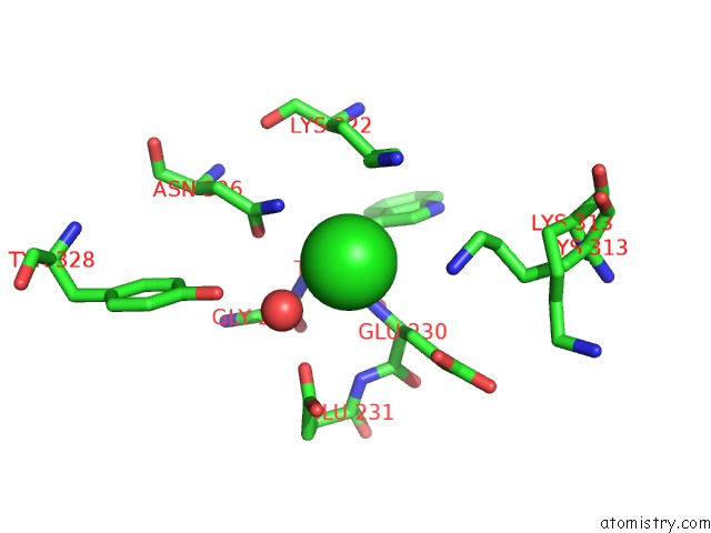
Mono view
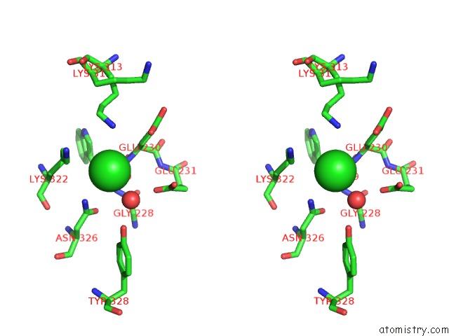
Stereo pair view
Mono view
Stereo pair view
A full contact list of Chlorine with other atoms in the Cl binding
site number 1 of Crystal Structure of A Putative Hydrolase (LPG1103) From Legionella Pneumophila Subsp. Pneumophila Str. Philadelphia 1 at 1.15 A Resolution within 5.0Å range:
|
Reference:
Joint Center For Structural Genomics (Jcsg),
Joint Center For Structural Genomics (Jcsg).
N/A N/A.
Page generated: Fri Jul 11 15:05:59 2025
Last articles
Mg in 4RNQMg in 4RO4
Mg in 4RO7
Mg in 4RNN
Mg in 4RNM
Mg in 4RNK
Mg in 4RNH
Mg in 4RN3
Mg in 4RL3
Mg in 4RML