Chlorine »
PDB 4fby-4fm6 »
4fex »
Chlorine in PDB 4fex: Crystal Structure of the Aminoglycoside Phosphotransferase Aph(3')-Ia, with Substrate Kanamycin and Small Molecule Inhibitor Tyrphostin AG1478
Enzymatic activity of Crystal Structure of the Aminoglycoside Phosphotransferase Aph(3')-Ia, with Substrate Kanamycin and Small Molecule Inhibitor Tyrphostin AG1478
All present enzymatic activity of Crystal Structure of the Aminoglycoside Phosphotransferase Aph(3')-Ia, with Substrate Kanamycin and Small Molecule Inhibitor Tyrphostin AG1478:
2.7.1.95;
2.7.1.95;
Protein crystallography data
The structure of Crystal Structure of the Aminoglycoside Phosphotransferase Aph(3')-Ia, with Substrate Kanamycin and Small Molecule Inhibitor Tyrphostin AG1478, PDB code: 4fex
was solved by
P.J.Stogios,
E.Evdokimova,
Z.Wawrzak,
G.Minasov,
O.Egorova,
R.Di Leo,
T.Shakya,
P.Spanogiannopoulos,
G.D.Wright,
A.Savchenko,
W.F.Anderson,
Center For Structural Genomics Of Infectious Diseases (Csgid),
with X-Ray Crystallography technique. A brief refinement statistics is given in the table below:
| Resolution Low / High (Å) | 19.90 / 2.71 |
| Space group | P 1 |
| Cell size a, b, c (Å), α, β, γ (°) | 57.669, 93.694, 96.318, 118.84, 103.56, 93.44 |
| R / Rfree (%) | 18.4 / 24 |
Other elements in 4fex:
The structure of Crystal Structure of the Aminoglycoside Phosphotransferase Aph(3')-Ia, with Substrate Kanamycin and Small Molecule Inhibitor Tyrphostin AG1478 also contains other interesting chemical elements:
| Sodium | (Na) | 2 atoms |
Chlorine Binding Sites:
The binding sites of Chlorine atom in the Crystal Structure of the Aminoglycoside Phosphotransferase Aph(3')-Ia, with Substrate Kanamycin and Small Molecule Inhibitor Tyrphostin AG1478
(pdb code 4fex). This binding sites where shown within
5.0 Angstroms radius around Chlorine atom.
In total 4 binding sites of Chlorine where determined in the Crystal Structure of the Aminoglycoside Phosphotransferase Aph(3')-Ia, with Substrate Kanamycin and Small Molecule Inhibitor Tyrphostin AG1478, PDB code: 4fex:
Jump to Chlorine binding site number: 1; 2; 3; 4;
In total 4 binding sites of Chlorine where determined in the Crystal Structure of the Aminoglycoside Phosphotransferase Aph(3')-Ia, with Substrate Kanamycin and Small Molecule Inhibitor Tyrphostin AG1478, PDB code: 4fex:
Jump to Chlorine binding site number: 1; 2; 3; 4;
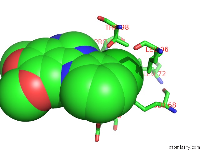
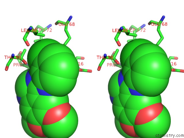
Chlorine binding site 1 out of 4 in 4fex
Go back to
Chlorine binding site 1 out
of 4 in the Crystal Structure of the Aminoglycoside Phosphotransferase Aph(3')-Ia, with Substrate Kanamycin and Small Molecule Inhibitor Tyrphostin AG1478
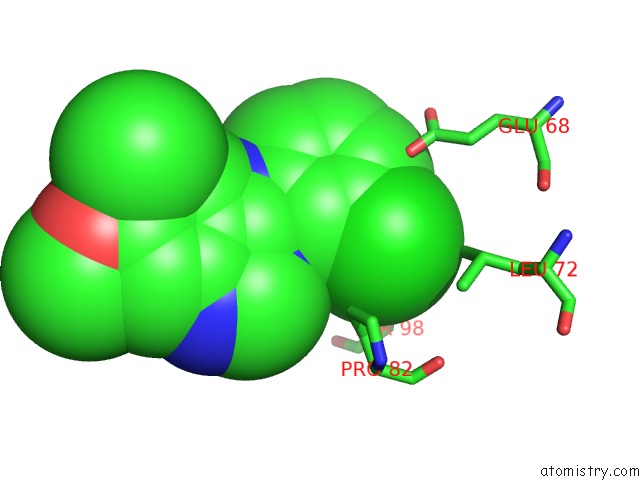
Mono view
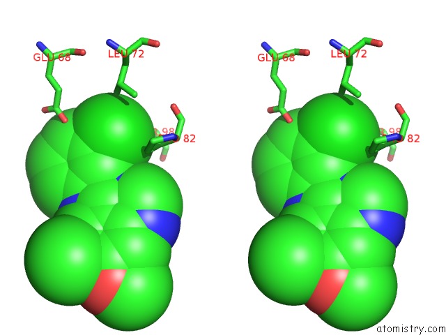
Stereo pair view
Mono view
Stereo pair view
A full contact list of Chlorine with other atoms in the Cl binding
site number 1 of Crystal Structure of the Aminoglycoside Phosphotransferase Aph(3')-Ia, with Substrate Kanamycin and Small Molecule Inhibitor Tyrphostin AG1478 within 5.0Å range:
|
Chlorine binding site 2 out of 4 in 4fex
Go back to
Chlorine binding site 2 out
of 4 in the Crystal Structure of the Aminoglycoside Phosphotransferase Aph(3')-Ia, with Substrate Kanamycin and Small Molecule Inhibitor Tyrphostin AG1478
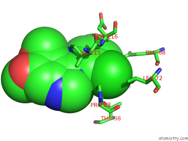
Mono view
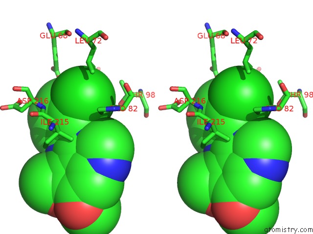
Stereo pair view
Mono view
Stereo pair view
A full contact list of Chlorine with other atoms in the Cl binding
site number 2 of Crystal Structure of the Aminoglycoside Phosphotransferase Aph(3')-Ia, with Substrate Kanamycin and Small Molecule Inhibitor Tyrphostin AG1478 within 5.0Å range:
|
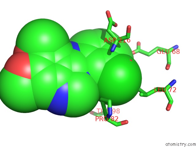
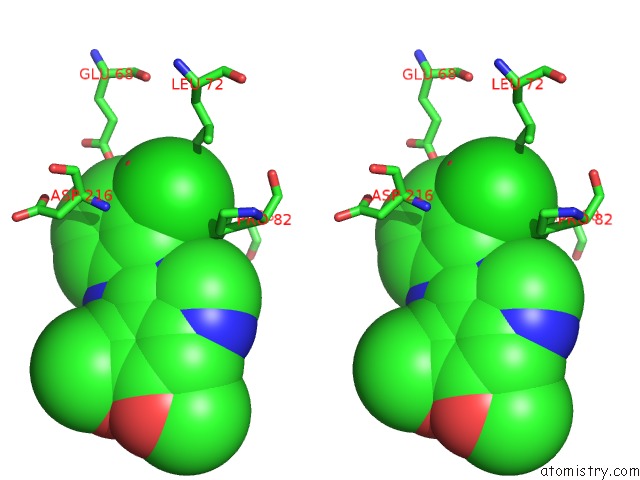
Chlorine binding site 3 out of 4 in 4fex
Go back to
Chlorine binding site 3 out
of 4 in the Crystal Structure of the Aminoglycoside Phosphotransferase Aph(3')-Ia, with Substrate Kanamycin and Small Molecule Inhibitor Tyrphostin AG1478
Mono view
Stereo pair view
Mono view
Stereo pair view
A full contact list of Chlorine with other atoms in the Cl binding
site number 3 of Crystal Structure of the Aminoglycoside Phosphotransferase Aph(3')-Ia, with Substrate Kanamycin and Small Molecule Inhibitor Tyrphostin AG1478 within 5.0Å range:
|
Chlorine binding site 4 out of 4 in 4fex
Go back to
Chlorine binding site 4 out
of 4 in the Crystal Structure of the Aminoglycoside Phosphotransferase Aph(3')-Ia, with Substrate Kanamycin and Small Molecule Inhibitor Tyrphostin AG1478
Mono view
Stereo pair view
Mono view
Stereo pair view
A full contact list of Chlorine with other atoms in the Cl binding
site number 4 of Crystal Structure of the Aminoglycoside Phosphotransferase Aph(3')-Ia, with Substrate Kanamycin and Small Molecule Inhibitor Tyrphostin AG1478 within 5.0Å range:
|
Reference:
P.J.Stogios,
P.Spanogiannopoulos,
E.Evdokimova,
O.Egorova,
T.Shakya,
N.Todorovic,
A.Capretta,
G.D.Wright,
A.Savchenko.
Structure-Guided Optimization of Protein Kinase Inhibitors Reverses Aminoglycoside Antibiotic Resistance. Biochem.J. V. 454 191 2013.
ISSN: ISSN 0264-6021
PubMed: 23758273
DOI: 10.1042/BJ20130317
Page generated: Fri Jul 11 15:11:55 2025
ISSN: ISSN 0264-6021
PubMed: 23758273
DOI: 10.1042/BJ20130317
Last articles
Mg in 4RI2Mg in 4RHX
Mg in 4RHU
Mg in 4RHT
Mg in 4RHD
Mg in 4RH7
Mg in 4RET
Mg in 4RGV
Mg in 4RGF
Mg in 4RF5