Chlorine »
PDB 4fv3-4g7y »
4g7v »
Chlorine in PDB 4g7v: Crystal Structure of Voltage Sensing Domain of Ci-Vsp with Fragment Antibody (R217E, 2.5 A)
Protein crystallography data
The structure of Crystal Structure of Voltage Sensing Domain of Ci-Vsp with Fragment Antibody (R217E, 2.5 A), PDB code: 4g7v
was solved by
Q.Li,
with X-Ray Crystallography technique. A brief refinement statistics is given in the table below:
| Resolution Low / High (Å) | 37.24 / 2.50 |
| Space group | P 65 2 2 |
| Cell size a, b, c (Å), α, β, γ (°) | 120.250, 120.250, 229.880, 90.00, 90.00, 120.00 |
| R / Rfree (%) | 20.1 / 24 |
Chlorine Binding Sites:
The binding sites of Chlorine atom in the Crystal Structure of Voltage Sensing Domain of Ci-Vsp with Fragment Antibody (R217E, 2.5 A)
(pdb code 4g7v). This binding sites where shown within
5.0 Angstroms radius around Chlorine atom.
In total only one binding site of Chlorine was determined in the Crystal Structure of Voltage Sensing Domain of Ci-Vsp with Fragment Antibody (R217E, 2.5 A), PDB code: 4g7v:
In total only one binding site of Chlorine was determined in the Crystal Structure of Voltage Sensing Domain of Ci-Vsp with Fragment Antibody (R217E, 2.5 A), PDB code: 4g7v:
Chlorine binding site 1 out of 1 in 4g7v
Go back to
Chlorine binding site 1 out
of 1 in the Crystal Structure of Voltage Sensing Domain of Ci-Vsp with Fragment Antibody (R217E, 2.5 A)
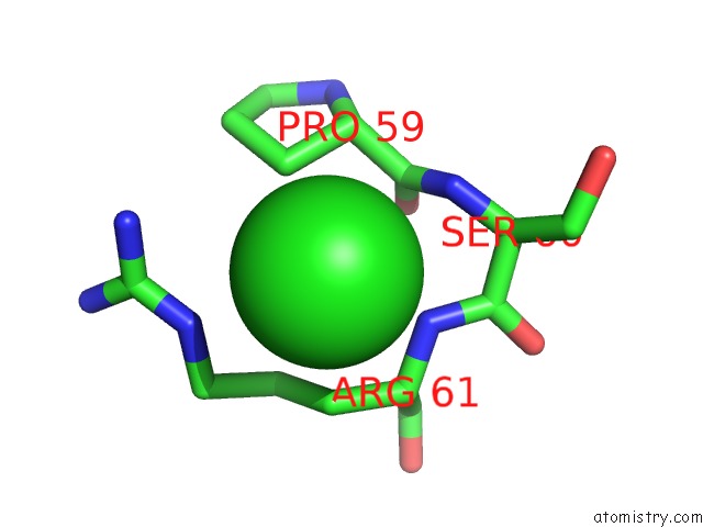
Mono view
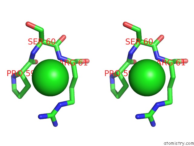
Stereo pair view
Mono view
Stereo pair view
A full contact list of Chlorine with other atoms in the Cl binding
site number 1 of Crystal Structure of Voltage Sensing Domain of Ci-Vsp with Fragment Antibody (R217E, 2.5 A) within 5.0Å range:
|
Reference:
Q.Li,
M.Paduch,
S.Wanderling,
R.E.Hulse,
S.Koide,
A.Kosiakoff,
E.Perozo.
Structural Rearrangement in Voltage Sensing Domain of Ci-Vsp To Be Published.
Page generated: Fri Jul 11 15:32:07 2025
Last articles
Mg in 4XC5Mg in 4XCL
Mg in 4XCJ
Mg in 4X8D
Mg in 4XA5
Mg in 4XC0
Mg in 4XBR
Mg in 4X9E
Mg in 4X8L
Mg in 4X8O