Chlorine »
PDB 4h1x-4h8x »
4h2f »
Chlorine in PDB 4h2f: Human Ecto-5'-Nucleotidase (CD73): Crystal Form I (Open) in Complex with Adenosine
Enzymatic activity of Human Ecto-5'-Nucleotidase (CD73): Crystal Form I (Open) in Complex with Adenosine
All present enzymatic activity of Human Ecto-5'-Nucleotidase (CD73): Crystal Form I (Open) in Complex with Adenosine:
3.1.3.5;
3.1.3.5;
Protein crystallography data
The structure of Human Ecto-5'-Nucleotidase (CD73): Crystal Form I (Open) in Complex with Adenosine, PDB code: 4h2f
was solved by
N.Straeter,
K.M.Knapp,
M.Zebisch,
J.Pippel,
with X-Ray Crystallography technique. A brief refinement statistics is given in the table below:
| Resolution Low / High (Å) | 41.89 / 1.85 |
| Space group | P 43 3 2 |
| Cell size a, b, c (Å), α, β, γ (°) | 167.548, 167.548, 167.548, 90.00, 90.00, 90.00 |
| R / Rfree (%) | 15.7 / 17.9 |
Other elements in 4h2f:
The structure of Human Ecto-5'-Nucleotidase (CD73): Crystal Form I (Open) in Complex with Adenosine also contains other interesting chemical elements:
| Calcium | (Ca) | 1 atom |
| Zinc | (Zn) | 1 atom |
Chlorine Binding Sites:
The binding sites of Chlorine atom in the Human Ecto-5'-Nucleotidase (CD73): Crystal Form I (Open) in Complex with Adenosine
(pdb code 4h2f). This binding sites where shown within
5.0 Angstroms radius around Chlorine atom.
In total only one binding site of Chlorine was determined in the Human Ecto-5'-Nucleotidase (CD73): Crystal Form I (Open) in Complex with Adenosine, PDB code: 4h2f:
In total only one binding site of Chlorine was determined in the Human Ecto-5'-Nucleotidase (CD73): Crystal Form I (Open) in Complex with Adenosine, PDB code: 4h2f:
Chlorine binding site 1 out of 1 in 4h2f
Go back to
Chlorine binding site 1 out
of 1 in the Human Ecto-5'-Nucleotidase (CD73): Crystal Form I (Open) in Complex with Adenosine
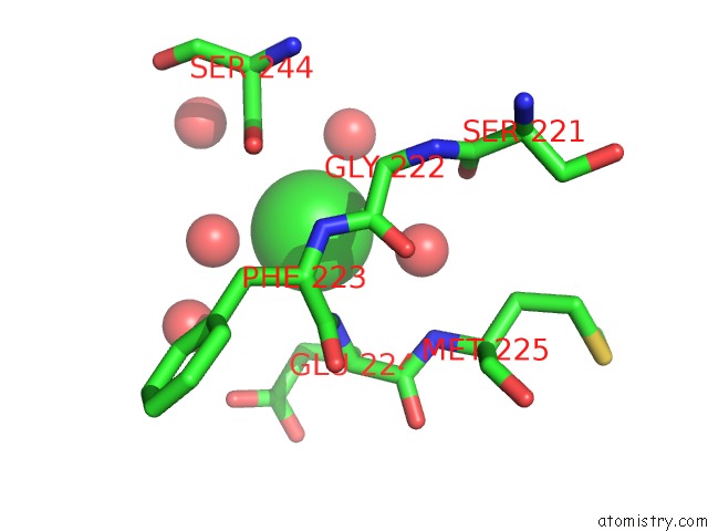
Mono view
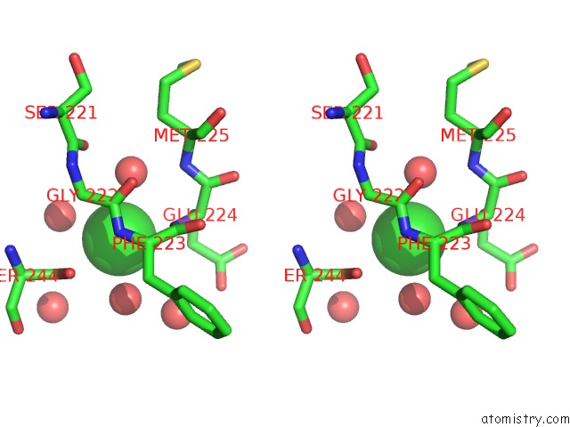
Stereo pair view
Mono view
Stereo pair view
A full contact list of Chlorine with other atoms in the Cl binding
site number 1 of Human Ecto-5'-Nucleotidase (CD73): Crystal Form I (Open) in Complex with Adenosine within 5.0Å range:
|
Reference:
K.Knapp,
M.Zebisch,
J.Pippel,
A.El-Tayeb,
C.E.Muller,
N.Strater.
Crystal Structure of the Human Ecto-5'-Nucleotidase (CD73): Insights Into the Regulation of Purinergic Signaling. Structure V. 20 2161 2012.
ISSN: ISSN 0969-2126
PubMed: 23142347
DOI: 10.1016/J.STR.2012.10.001
Page generated: Fri Jul 11 16:06:43 2025
ISSN: ISSN 0969-2126
PubMed: 23142347
DOI: 10.1016/J.STR.2012.10.001
Last articles
Mg in 4CT4Mg in 4CVM
Mg in 4CVO
Mg in 4CVL
Mg in 4CVH
Mg in 4CTA
Mg in 4CS4
Mg in 4CS3
Mg in 4CS1
Mg in 4CS0