Chlorine »
PDB 4hn0-4hw2 »
4hus »
Chlorine in PDB 4hus: Crystal Structure of Streptogramin Group A Antibiotic Acetyltransferase Vata From Staphylococcus Aureus in Complex with Virginiamycin M1
Protein crystallography data
The structure of Crystal Structure of Streptogramin Group A Antibiotic Acetyltransferase Vata From Staphylococcus Aureus in Complex with Virginiamycin M1, PDB code: 4hus
was solved by
P.J.Stogios,
G.Minasov,
E.Evdokimova,
Z.Wawrzak,
V.Yim,
M.Krishnamoorthy,
R.Di Leo,
P.Courvalin,
A.Savchenko,
W.F.Anderson,
Center For Structuralgenomics Of Infectious Diseases (Csgid),
with X-Ray Crystallography technique. A brief refinement statistics is given in the table below:
| Resolution Low / High (Å) | 29.39 / 2.36 |
| Space group | C 2 2 21 |
| Cell size a, b, c (Å), α, β, γ (°) | 93.308, 184.735, 98.567, 90.00, 90.00, 90.00 |
| R / Rfree (%) | 18.4 / 22.8 |
Other elements in 4hus:
The structure of Crystal Structure of Streptogramin Group A Antibiotic Acetyltransferase Vata From Staphylococcus Aureus in Complex with Virginiamycin M1 also contains other interesting chemical elements:
| Sodium | (Na) | 1 atom |
Chlorine Binding Sites:
The binding sites of Chlorine atom in the Crystal Structure of Streptogramin Group A Antibiotic Acetyltransferase Vata From Staphylococcus Aureus in Complex with Virginiamycin M1
(pdb code 4hus). This binding sites where shown within
5.0 Angstroms radius around Chlorine atom.
In total 9 binding sites of Chlorine where determined in the Crystal Structure of Streptogramin Group A Antibiotic Acetyltransferase Vata From Staphylococcus Aureus in Complex with Virginiamycin M1, PDB code: 4hus:
Jump to Chlorine binding site number: 1; 2; 3; 4; 5; 6; 7; 8; 9;
In total 9 binding sites of Chlorine where determined in the Crystal Structure of Streptogramin Group A Antibiotic Acetyltransferase Vata From Staphylococcus Aureus in Complex with Virginiamycin M1, PDB code: 4hus:
Jump to Chlorine binding site number: 1; 2; 3; 4; 5; 6; 7; 8; 9;
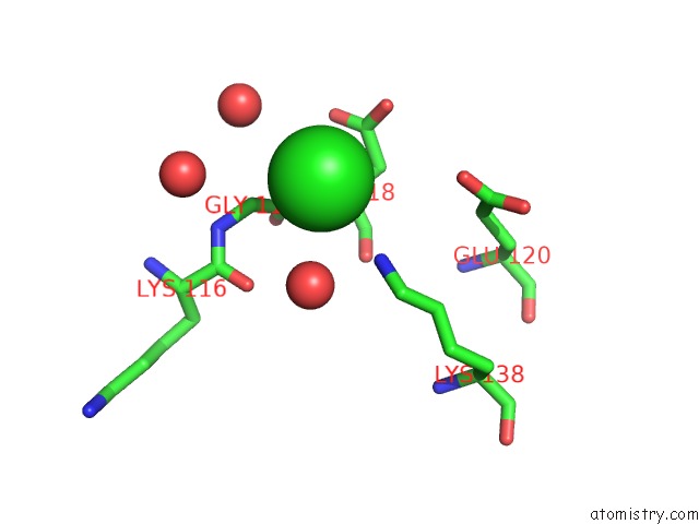
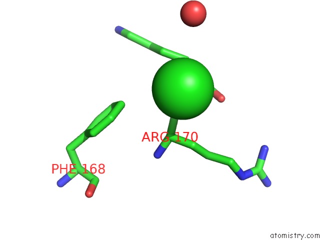
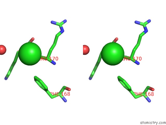
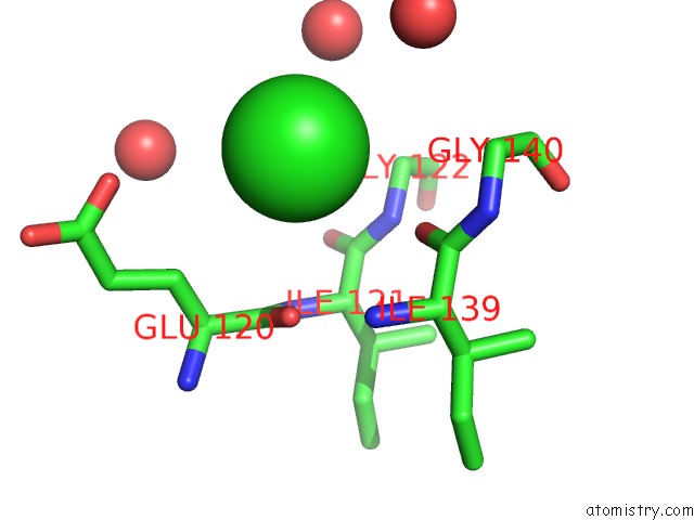
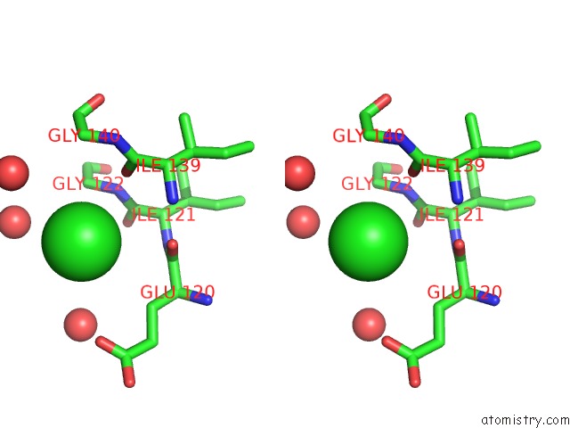
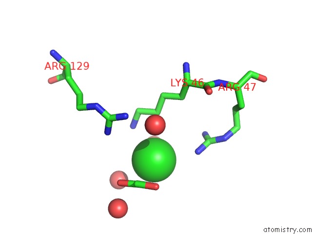
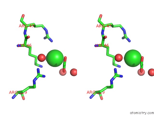
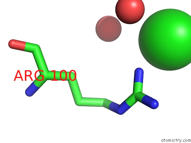
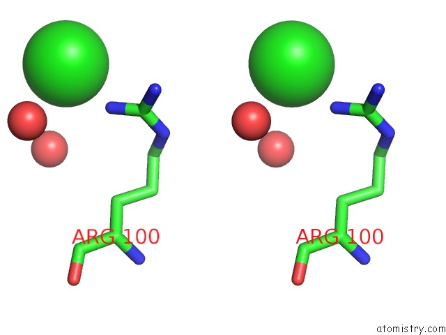
Chlorine binding site 1 out of 9 in 4hus
Go back to
Chlorine binding site 1 out
of 9 in the Crystal Structure of Streptogramin Group A Antibiotic Acetyltransferase Vata From Staphylococcus Aureus in Complex with Virginiamycin M1
Mono view
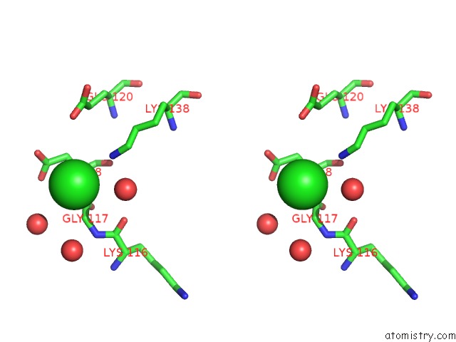
Stereo pair view
Mono view
Stereo pair view
A full contact list of Chlorine with other atoms in the Cl binding
site number 1 of Crystal Structure of Streptogramin Group A Antibiotic Acetyltransferase Vata From Staphylococcus Aureus in Complex with Virginiamycin M1 within 5.0Å range:
|
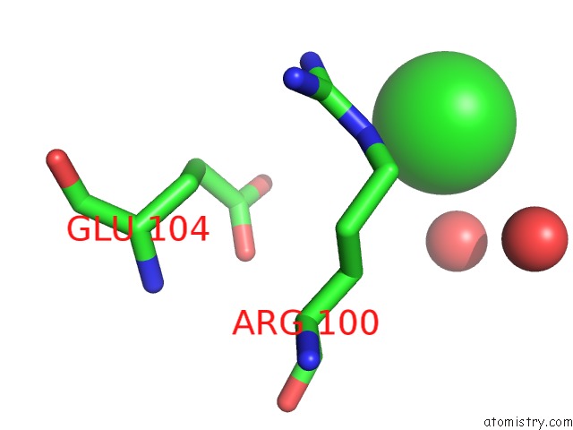
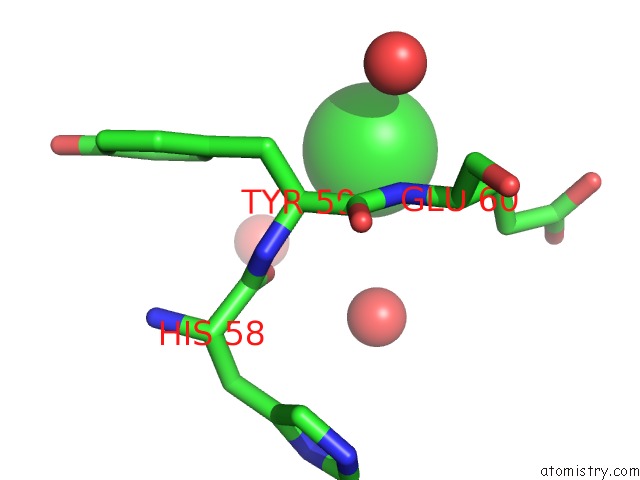
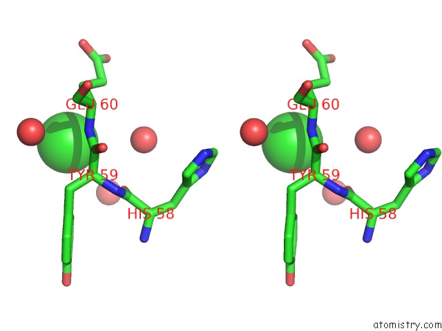
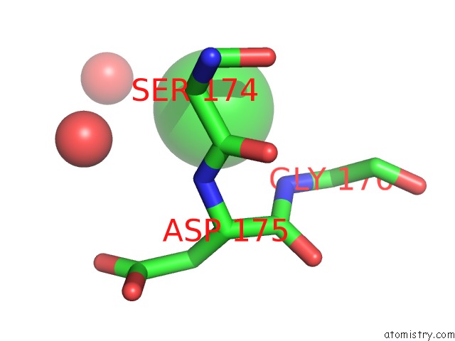
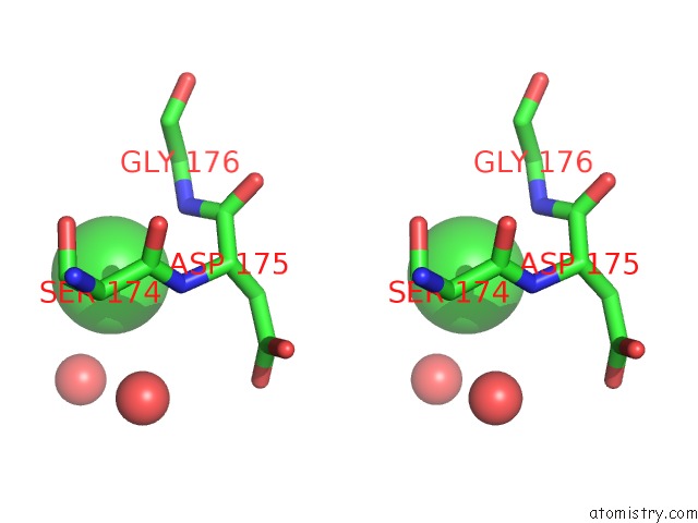
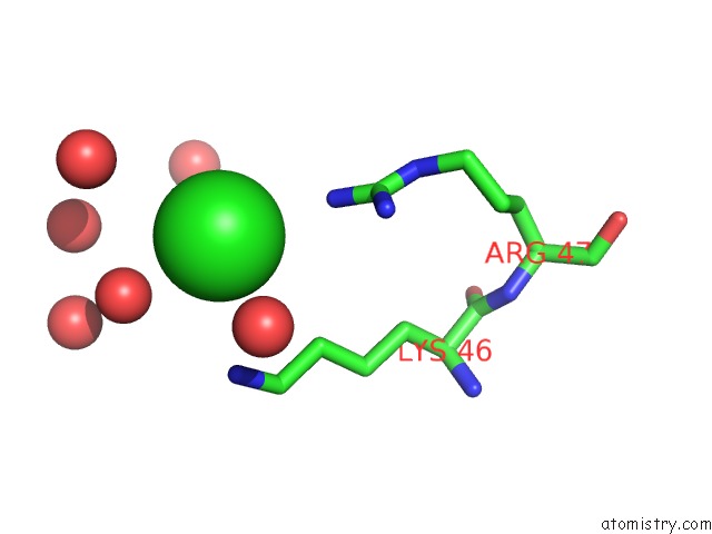
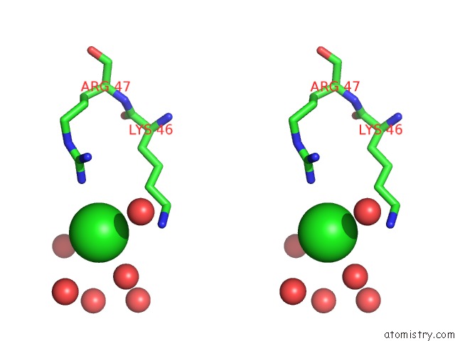
Chlorine binding site 2 out of 9 in 4hus
Go back to
Chlorine binding site 2 out
of 9 in the Crystal Structure of Streptogramin Group A Antibiotic Acetyltransferase Vata From Staphylococcus Aureus in Complex with Virginiamycin M1
Mono view
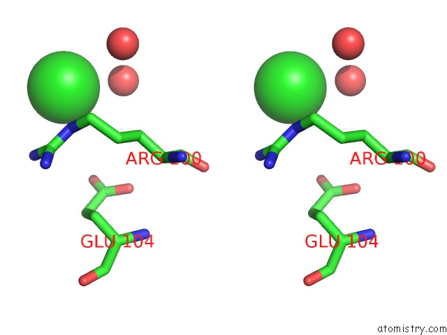
Stereo pair view
Mono view
Stereo pair view
A full contact list of Chlorine with other atoms in the Cl binding
site number 2 of Crystal Structure of Streptogramin Group A Antibiotic Acetyltransferase Vata From Staphylococcus Aureus in Complex with Virginiamycin M1 within 5.0Å range:
|
Chlorine binding site 3 out of 9 in 4hus
Go back to
Chlorine binding site 3 out
of 9 in the Crystal Structure of Streptogramin Group A Antibiotic Acetyltransferase Vata From Staphylococcus Aureus in Complex with Virginiamycin M1
Mono view
Stereo pair view
Mono view
Stereo pair view
A full contact list of Chlorine with other atoms in the Cl binding
site number 3 of Crystal Structure of Streptogramin Group A Antibiotic Acetyltransferase Vata From Staphylococcus Aureus in Complex with Virginiamycin M1 within 5.0Å range:
|
Chlorine binding site 4 out of 9 in 4hus
Go back to
Chlorine binding site 4 out
of 9 in the Crystal Structure of Streptogramin Group A Antibiotic Acetyltransferase Vata From Staphylococcus Aureus in Complex with Virginiamycin M1
Mono view
Stereo pair view
Mono view
Stereo pair view
A full contact list of Chlorine with other atoms in the Cl binding
site number 4 of Crystal Structure of Streptogramin Group A Antibiotic Acetyltransferase Vata From Staphylococcus Aureus in Complex with Virginiamycin M1 within 5.0Å range:
|
Chlorine binding site 5 out of 9 in 4hus
Go back to
Chlorine binding site 5 out
of 9 in the Crystal Structure of Streptogramin Group A Antibiotic Acetyltransferase Vata From Staphylococcus Aureus in Complex with Virginiamycin M1
Mono view
Stereo pair view
Mono view
Stereo pair view
A full contact list of Chlorine with other atoms in the Cl binding
site number 5 of Crystal Structure of Streptogramin Group A Antibiotic Acetyltransferase Vata From Staphylococcus Aureus in Complex with Virginiamycin M1 within 5.0Å range:
|
Chlorine binding site 6 out of 9 in 4hus
Go back to
Chlorine binding site 6 out
of 9 in the Crystal Structure of Streptogramin Group A Antibiotic Acetyltransferase Vata From Staphylococcus Aureus in Complex with Virginiamycin M1
Mono view
Stereo pair view
Mono view
Stereo pair view
A full contact list of Chlorine with other atoms in the Cl binding
site number 6 of Crystal Structure of Streptogramin Group A Antibiotic Acetyltransferase Vata From Staphylococcus Aureus in Complex with Virginiamycin M1 within 5.0Å range:
|
Chlorine binding site 7 out of 9 in 4hus
Go back to
Chlorine binding site 7 out
of 9 in the Crystal Structure of Streptogramin Group A Antibiotic Acetyltransferase Vata From Staphylococcus Aureus in Complex with Virginiamycin M1
Mono view
Stereo pair view
Mono view
Stereo pair view
A full contact list of Chlorine with other atoms in the Cl binding
site number 7 of Crystal Structure of Streptogramin Group A Antibiotic Acetyltransferase Vata From Staphylococcus Aureus in Complex with Virginiamycin M1 within 5.0Å range:
|
Chlorine binding site 8 out of 9 in 4hus
Go back to
Chlorine binding site 8 out
of 9 in the Crystal Structure of Streptogramin Group A Antibiotic Acetyltransferase Vata From Staphylococcus Aureus in Complex with Virginiamycin M1
Mono view
Stereo pair view
Mono view
Stereo pair view
A full contact list of Chlorine with other atoms in the Cl binding
site number 8 of Crystal Structure of Streptogramin Group A Antibiotic Acetyltransferase Vata From Staphylococcus Aureus in Complex with Virginiamycin M1 within 5.0Å range:
|
Chlorine binding site 9 out of 9 in 4hus
Go back to
Chlorine binding site 9 out
of 9 in the Crystal Structure of Streptogramin Group A Antibiotic Acetyltransferase Vata From Staphylococcus Aureus in Complex with Virginiamycin M1
Mono view
Stereo pair view
Mono view
Stereo pair view
A full contact list of Chlorine with other atoms in the Cl binding
site number 9 of Crystal Structure of Streptogramin Group A Antibiotic Acetyltransferase Vata From Staphylococcus Aureus in Complex with Virginiamycin M1 within 5.0Å range:
|
Reference:
P.J.Stogios,
M.L.Kuhn,
E.Evdokimova,
P.Courvalin,
W.F.Anderson,
A.Savchenko.
Potential For Reduction of Streptogramin A Resistance Revealed By Structural Analysis of Acetyltransferase Vata. Antimicrob.Agents Chemother. V. 58 7083 2014.
ISSN: ISSN 0066-4804
PubMed: 25223995
DOI: 10.1128/AAC.03743-14
Page generated: Fri Jul 11 16:32:19 2025
ISSN: ISSN 0066-4804
PubMed: 25223995
DOI: 10.1128/AAC.03743-14
Last articles
Mg in 5M8GMg in 5M8D
Mg in 5M8B
Mg in 5M7Y
Mg in 5M7Q
Mg in 5M7P
Mg in 5M7L
Mg in 5M7N
Mg in 5M7O
Mg in 5M7K