Chlorine »
PDB 4ibf-4iij »
4ihj »
Chlorine in PDB 4ihj: Crystal Structure of Tubulin-Stathmin-Ttl-Adp Complex
Protein crystallography data
The structure of Crystal Structure of Tubulin-Stathmin-Ttl-Adp Complex, PDB code: 4ihj
was solved by
A.E.Prota,
M.M.Magiera,
M.Kuijpers,
K.Bargsten,
D.Frey,
M.Wieser,
R.Jaussi,
C.C.Hoogenraad,
R.A.Kammerer,
C.Janke,
M.O.Steinmetz,
with X-Ray Crystallography technique. A brief refinement statistics is given in the table below:
| Resolution Low / High (Å) | 72.10 / 2.00 |
| Space group | P 21 21 21 |
| Cell size a, b, c (Å), α, β, γ (°) | 104.520, 157.310, 180.990, 90.00, 90.00, 90.00 |
| R / Rfree (%) | 16.8 / 21.7 |
Other elements in 4ihj:
The structure of Crystal Structure of Tubulin-Stathmin-Ttl-Adp Complex also contains other interesting chemical elements:
| Magnesium | (Mg) | 7 atoms |
| Calcium | (Ca) | 4 atoms |
Chlorine Binding Sites:
The binding sites of Chlorine atom in the Crystal Structure of Tubulin-Stathmin-Ttl-Adp Complex
(pdb code 4ihj). This binding sites where shown within
5.0 Angstroms radius around Chlorine atom.
In total only one binding site of Chlorine was determined in the Crystal Structure of Tubulin-Stathmin-Ttl-Adp Complex, PDB code: 4ihj:
In total only one binding site of Chlorine was determined in the Crystal Structure of Tubulin-Stathmin-Ttl-Adp Complex, PDB code: 4ihj:
Chlorine binding site 1 out of 1 in 4ihj
Go back to
Chlorine binding site 1 out
of 1 in the Crystal Structure of Tubulin-Stathmin-Ttl-Adp Complex
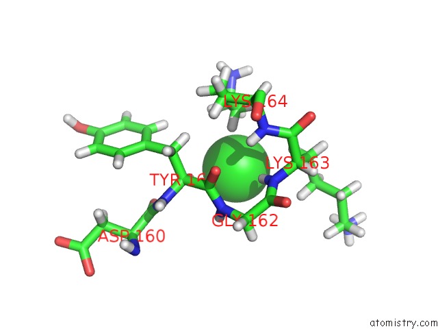
Mono view
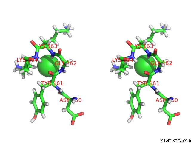
Stereo pair view
Mono view
Stereo pair view
A full contact list of Chlorine with other atoms in the Cl binding
site number 1 of Crystal Structure of Tubulin-Stathmin-Ttl-Adp Complex within 5.0Å range:
|
Reference:
A.E.Prota,
M.M.Magiera,
M.Kuijpers,
K.Bargsten,
D.Frey,
M.Wieser,
R.Jaussi,
C.C.Hoogenraad,
R.A.Kammerer,
C.Janke,
M.O.Steinmetz.
Structural Basis of Tubulin Tyrosination By Tubulin Tyrosine Ligase. J.Cell Biol. V. 200 259 2013.
ISSN: ISSN 0021-9525
PubMed: 23358242
DOI: 10.1083/JCB.201211017
Page generated: Fri Jul 11 16:50:41 2025
ISSN: ISSN 0021-9525
PubMed: 23358242
DOI: 10.1083/JCB.201211017
Last articles
Mg in 3A12Mg in 3A3Y
Mg in 3A13
Mg in 3A1U
Mg in 3A1S
Mg in 3A1D
Mg in 3A1E
Mg in 3A16
Mg in 3A1C
Mg in 3A14