Chlorine »
PDB 4j93-4jk9 »
4jec »
Chlorine in PDB 4jec: Joint Neutron and X-Ray Structure of Per-Deuterated Hiv-1 Protease in Complex with Clinical Inhibitor Amprenavir
Enzymatic activity of Joint Neutron and X-Ray Structure of Per-Deuterated Hiv-1 Protease in Complex with Clinical Inhibitor Amprenavir
All present enzymatic activity of Joint Neutron and X-Ray Structure of Per-Deuterated Hiv-1 Protease in Complex with Clinical Inhibitor Amprenavir:
3.4.23.16;
3.4.23.16;
Protein crystallography data
The structure of Joint Neutron and X-Ray Structure of Per-Deuterated Hiv-1 Protease in Complex with Clinical Inhibitor Amprenavir, PDB code: 4jec
was solved by
A.Y.Kovalevsky,
I.T.Weber,
P.Langan,
with X-Ray Crystallography technique. A brief refinement statistics is given in the table below:
| Resolution Low / High (Å) | N/A / 2.01 |
| Space group | P 21 21 2 |
| Cell size a, b, c (Å), α, β, γ (°) | 59.186, 87.431, 46.405, 90.00, 90.00, 90.00 |
| R / Rfree (%) | 24.4 / 26.1 |
Chlorine Binding Sites:
The binding sites of Chlorine atom in the Joint Neutron and X-Ray Structure of Per-Deuterated Hiv-1 Protease in Complex with Clinical Inhibitor Amprenavir
(pdb code 4jec). This binding sites where shown within
5.0 Angstroms radius around Chlorine atom.
In total only one binding site of Chlorine was determined in the Joint Neutron and X-Ray Structure of Per-Deuterated Hiv-1 Protease in Complex with Clinical Inhibitor Amprenavir, PDB code: 4jec:
In total only one binding site of Chlorine was determined in the Joint Neutron and X-Ray Structure of Per-Deuterated Hiv-1 Protease in Complex with Clinical Inhibitor Amprenavir, PDB code: 4jec:
Chlorine binding site 1 out of 1 in 4jec
Go back to
Chlorine binding site 1 out
of 1 in the Joint Neutron and X-Ray Structure of Per-Deuterated Hiv-1 Protease in Complex with Clinical Inhibitor Amprenavir
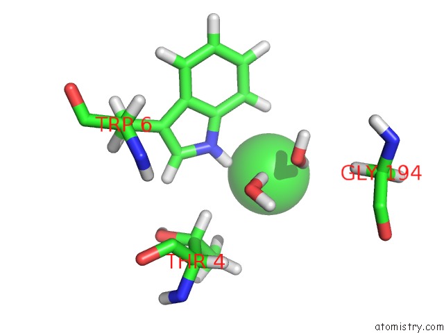
Mono view
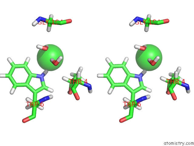
Stereo pair view
Mono view
Stereo pair view
A full contact list of Chlorine with other atoms in the Cl binding
site number 1 of Joint Neutron and X-Ray Structure of Per-Deuterated Hiv-1 Protease in Complex with Clinical Inhibitor Amprenavir within 5.0Å range:
|
Reference:
I.T.Weber,
M.J.Waltman,
M.Mustyakimov,
M.P.Blakeley,
D.A.Keen,
A.K.Ghosh,
P.Langan,
A.Y.Kovalevsky.
Joint X-Ray/Neutron Crystallographic Study of Hiv-1 Protease with Clinical Inhibitor Amprenavir: Insights For Drug Design. J.Med.Chem. V. 56 5631 2013.
ISSN: ISSN 0022-2623
PubMed: 23772563
DOI: 10.1021/JM400684F
Page generated: Fri Jul 11 17:17:59 2025
ISSN: ISSN 0022-2623
PubMed: 23772563
DOI: 10.1021/JM400684F
Last articles
Mg in 6U1EMg in 6U1D
Mg in 6TXA
Mg in 6U0U
Mg in 6U0T
Mg in 6TXC
Mg in 6U0H
Mg in 6U01
Mg in 6U07
Mg in 6U05