Chlorine »
PDB 4k2f-4kbb »
4k6c »
Chlorine in PDB 4k6c: X-Ray Crystal Structure of A Putative Acetoacyl-Coa Reductase From Burkholderia Cenocepacia
Enzymatic activity of X-Ray Crystal Structure of A Putative Acetoacyl-Coa Reductase From Burkholderia Cenocepacia
All present enzymatic activity of X-Ray Crystal Structure of A Putative Acetoacyl-Coa Reductase From Burkholderia Cenocepacia:
1.1.1.36;
1.1.1.36;
Protein crystallography data
The structure of X-Ray Crystal Structure of A Putative Acetoacyl-Coa Reductase From Burkholderia Cenocepacia, PDB code: 4k6c
was solved by
Seattle Structural Genomics Center For Infectious Disease (Ssgcid),
with X-Ray Crystallography technique. A brief refinement statistics is given in the table below:
| Resolution Low / High (Å) | 42.28 / 1.85 |
| Space group | P 31 2 1 |
| Cell size a, b, c (Å), α, β, γ (°) | 84.480, 84.480, 143.160, 90.00, 90.00, 120.00 |
| R / Rfree (%) | 16 / 19.6 |
Chlorine Binding Sites:
The binding sites of Chlorine atom in the X-Ray Crystal Structure of A Putative Acetoacyl-Coa Reductase From Burkholderia Cenocepacia
(pdb code 4k6c). This binding sites where shown within
5.0 Angstroms radius around Chlorine atom.
In total only one binding site of Chlorine was determined in the X-Ray Crystal Structure of A Putative Acetoacyl-Coa Reductase From Burkholderia Cenocepacia, PDB code: 4k6c:
In total only one binding site of Chlorine was determined in the X-Ray Crystal Structure of A Putative Acetoacyl-Coa Reductase From Burkholderia Cenocepacia, PDB code: 4k6c:
Chlorine binding site 1 out of 1 in 4k6c
Go back to
Chlorine binding site 1 out
of 1 in the X-Ray Crystal Structure of A Putative Acetoacyl-Coa Reductase From Burkholderia Cenocepacia
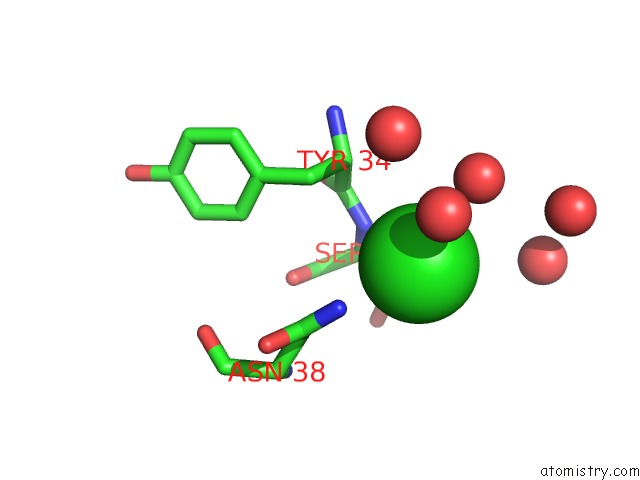
Mono view
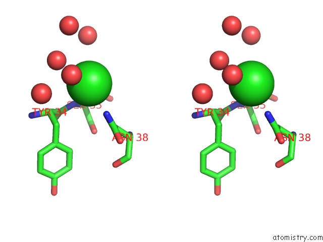
Stereo pair view
Mono view
Stereo pair view
A full contact list of Chlorine with other atoms in the Cl binding
site number 1 of X-Ray Crystal Structure of A Putative Acetoacyl-Coa Reductase From Burkholderia Cenocepacia within 5.0Å range:
|
Reference:
J.W.Fairman,
T.E.Edwards,
D.Lorimer.
X-Ray Crystal Structure of A Putative Acetoacyl-Coa Reductase From Burkholderia Cenocepacia To Be Published.
Page generated: Fri Jul 11 17:40:08 2025
Last articles
Mg in 5R8UMg in 5QTQ
Mg in 5QTP
Mg in 5QTO
Mg in 5QTN
Mg in 5QTM
Mg in 5QTL
Mg in 5QKB
Mg in 5QQC
Mg in 5QKA