Chlorine »
PDB 4k2f-4kbb »
4k7v »
Chlorine in PDB 4k7v: OYE1-W116A Complexed with (R)-Carvone
Enzymatic activity of OYE1-W116A Complexed with (R)-Carvone
All present enzymatic activity of OYE1-W116A Complexed with (R)-Carvone:
1.6.99.1;
1.6.99.1;
Protein crystallography data
The structure of OYE1-W116A Complexed with (R)-Carvone, PDB code: 4k7v
was solved by
B.Sullivan,
Y.A.Pompeu,
J.D.Stewart,
with X-Ray Crystallography technique. A brief refinement statistics is given in the table below:
| Resolution Low / High (Å) | 35.42 / 1.52 |
| Space group | P 43 21 2 |
| Cell size a, b, c (Å), α, β, γ (°) | 140.853, 140.853, 42.841, 90.00, 90.00, 90.00 |
| R / Rfree (%) | 14.5 / 18.3 |
Other elements in 4k7v:
The structure of OYE1-W116A Complexed with (R)-Carvone also contains other interesting chemical elements:
| Magnesium | (Mg) | 1 atom |
| Sodium | (Na) | 2 atoms |
Chlorine Binding Sites:
The binding sites of Chlorine atom in the OYE1-W116A Complexed with (R)-Carvone
(pdb code 4k7v). This binding sites where shown within
5.0 Angstroms radius around Chlorine atom.
In total only one binding site of Chlorine was determined in the OYE1-W116A Complexed with (R)-Carvone, PDB code: 4k7v:
In total only one binding site of Chlorine was determined in the OYE1-W116A Complexed with (R)-Carvone, PDB code: 4k7v:
Chlorine binding site 1 out of 1 in 4k7v
Go back to
Chlorine binding site 1 out
of 1 in the OYE1-W116A Complexed with (R)-Carvone
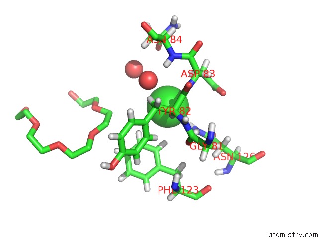
Mono view
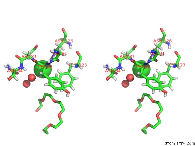
Stereo pair view
Mono view
Stereo pair view
A full contact list of Chlorine with other atoms in the Cl binding
site number 1 of OYE1-W116A Complexed with (R)-Carvone within 5.0Å range:
|
Reference:
Y.A.Pompeu,
B.Sullivan,
J.D.Stewart.
X‑Ray Crystallography Reveals How Subtle Changes Control the Orientation of Substrate Binding in An Alkene Reductase Acs Catalysis V. 3 2376 2013.
ISSN: ESSN 2155-5435
DOI: 10.1021/CS400622E
Page generated: Fri Jul 11 17:41:19 2025
ISSN: ESSN 2155-5435
DOI: 10.1021/CS400622E
Last articles
Mg in 5SEEMg in 5SEC
Mg in 5SEB
Mg in 5SEA
Mg in 5SE9
Mg in 5SE8
Mg in 5SE7
Mg in 5SE6
Mg in 5SE5
Mg in 5SE4