Chlorine »
PDB 4kbn-4kmz »
4kge »
Chlorine in PDB 4kge: Crystal Structure of Near-Infrared Fluorescent Protein with An Extended Stokes Shift, pH 4.5
Protein crystallography data
The structure of Crystal Structure of Near-Infrared Fluorescent Protein with An Extended Stokes Shift, pH 4.5, PDB code: 4kge
was solved by
V.N.Malashkevich,
K.Piatkevich,
S.C.Almo,
V.Verkhusha,
New Yorkstructural Genomics Research Consortium (Nysgrc),
with X-Ray Crystallography technique. A brief refinement statistics is given in the table below:
| Resolution Low / High (Å) | 47.11 / 2.30 |
| Space group | P 61 2 2 |
| Cell size a, b, c (Å), α, β, γ (°) | 106.050, 106.050, 219.526, 90.00, 90.00, 120.00 |
| R / Rfree (%) | 18.8 / 23.2 |
Chlorine Binding Sites:
The binding sites of Chlorine atom in the Crystal Structure of Near-Infrared Fluorescent Protein with An Extended Stokes Shift, pH 4.5
(pdb code 4kge). This binding sites where shown within
5.0 Angstroms radius around Chlorine atom.
In total 3 binding sites of Chlorine where determined in the Crystal Structure of Near-Infrared Fluorescent Protein with An Extended Stokes Shift, pH 4.5, PDB code: 4kge:
Jump to Chlorine binding site number: 1; 2; 3;
In total 3 binding sites of Chlorine where determined in the Crystal Structure of Near-Infrared Fluorescent Protein with An Extended Stokes Shift, pH 4.5, PDB code: 4kge:
Jump to Chlorine binding site number: 1; 2; 3;
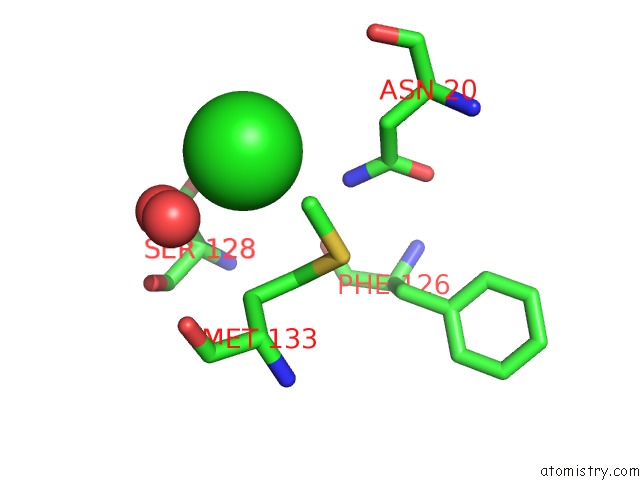
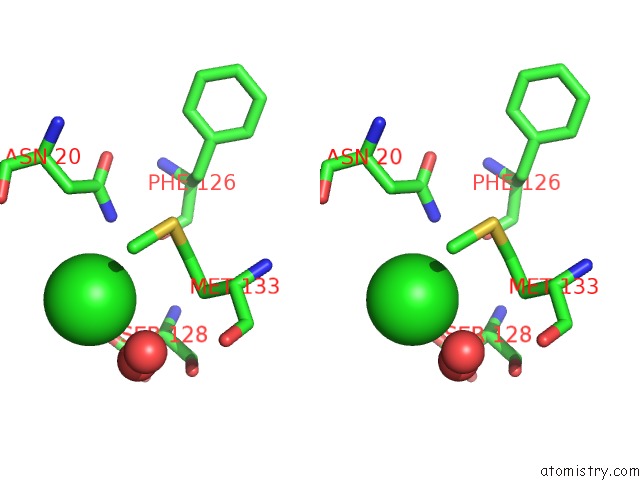
Chlorine binding site 1 out of 3 in 4kge
Go back to
Chlorine binding site 1 out
of 3 in the Crystal Structure of Near-Infrared Fluorescent Protein with An Extended Stokes Shift, pH 4.5
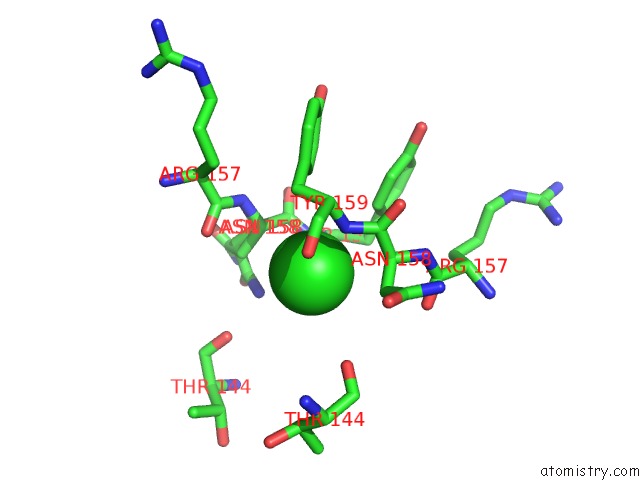
Mono view
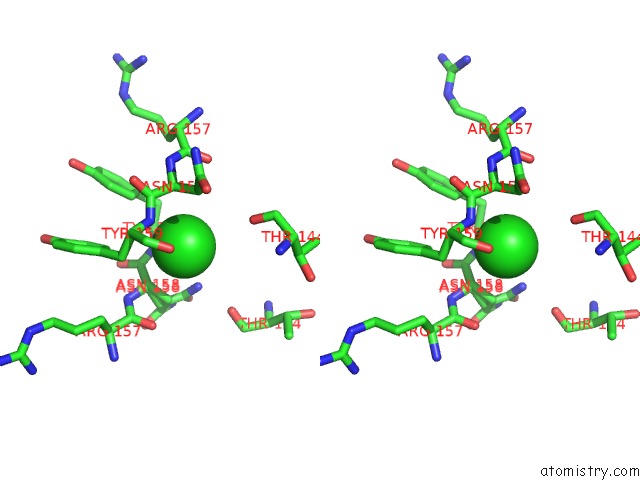
Stereo pair view
Mono view
Stereo pair view
A full contact list of Chlorine with other atoms in the Cl binding
site number 1 of Crystal Structure of Near-Infrared Fluorescent Protein with An Extended Stokes Shift, pH 4.5 within 5.0Å range:
|
Chlorine binding site 2 out of 3 in 4kge
Go back to
Chlorine binding site 2 out
of 3 in the Crystal Structure of Near-Infrared Fluorescent Protein with An Extended Stokes Shift, pH 4.5
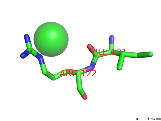
Mono view
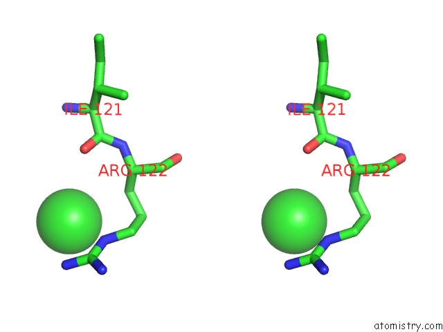
Stereo pair view
Mono view
Stereo pair view
A full contact list of Chlorine with other atoms in the Cl binding
site number 2 of Crystal Structure of Near-Infrared Fluorescent Protein with An Extended Stokes Shift, pH 4.5 within 5.0Å range:
|
Chlorine binding site 3 out of 3 in 4kge
Go back to
Chlorine binding site 3 out
of 3 in the Crystal Structure of Near-Infrared Fluorescent Protein with An Extended Stokes Shift, pH 4.5
Mono view
Stereo pair view
Mono view
Stereo pair view
A full contact list of Chlorine with other atoms in the Cl binding
site number 3 of Crystal Structure of Near-Infrared Fluorescent Protein with An Extended Stokes Shift, pH 4.5 within 5.0Å range:
|
Reference:
K.D.Piatkevich,
V.N.Malashkevich,
K.S.Morozova,
N.A.Nemkovich,
S.C.Almo,
V.V.Verkhusha.
Extended Stokes Shift in Fluorescent Proteins: Chromophore-Protein Interactions in A Near-Infrared TAGRFP675 Variant. Sci Rep V. 3 1847 2013.
ISSN: ESSN 2045-2322
PubMed: 23677204
DOI: 10.1038/SREP01847
Page generated: Fri Jul 11 17:45:04 2025
ISSN: ESSN 2045-2322
PubMed: 23677204
DOI: 10.1038/SREP01847
Last articles
Mg in 3SUCMg in 3ST8
Mg in 3SU8
Mg in 3STP
Mg in 3SSM
Mg in 3SSN
Mg in 3SSF
Mg in 3SS8
Mg in 3SRF
Mg in 3SRD