Chlorine »
PDB 4kn0-4kwt »
4kqo »
Chlorine in PDB 4kqo: Crystal Structure of Penicillin-Binding Protein 3 From Pseudomonas Aeruginosa in Complex with Piperacillin
Protein crystallography data
The structure of Crystal Structure of Penicillin-Binding Protein 3 From Pseudomonas Aeruginosa in Complex with Piperacillin, PDB code: 4kqo
was solved by
J.E.Nettleship,
D.I.Stuart,
R.J.Owens,
J.Ren,
with X-Ray Crystallography technique. A brief refinement statistics is given in the table below:
| Resolution Low / High (Å) | 47.10 / 2.31 |
| Space group | P 1 |
| Cell size a, b, c (Å), α, β, γ (°) | 57.049, 74.935, 82.785, 71.26, 86.04, 85.59 |
| R / Rfree (%) | 21.7 / 25 |
Chlorine Binding Sites:
The binding sites of Chlorine atom in the Crystal Structure of Penicillin-Binding Protein 3 From Pseudomonas Aeruginosa in Complex with Piperacillin
(pdb code 4kqo). This binding sites where shown within
5.0 Angstroms radius around Chlorine atom.
In total 2 binding sites of Chlorine where determined in the Crystal Structure of Penicillin-Binding Protein 3 From Pseudomonas Aeruginosa in Complex with Piperacillin, PDB code: 4kqo:
Jump to Chlorine binding site number: 1; 2;
In total 2 binding sites of Chlorine where determined in the Crystal Structure of Penicillin-Binding Protein 3 From Pseudomonas Aeruginosa in Complex with Piperacillin, PDB code: 4kqo:
Jump to Chlorine binding site number: 1; 2;
Chlorine binding site 1 out of 2 in 4kqo
Go back to
Chlorine binding site 1 out
of 2 in the Crystal Structure of Penicillin-Binding Protein 3 From Pseudomonas Aeruginosa in Complex with Piperacillin
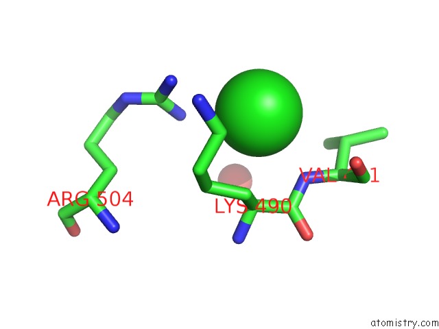
Mono view
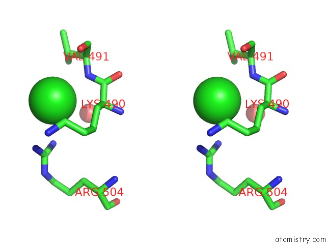
Stereo pair view
Mono view
Stereo pair view
A full contact list of Chlorine with other atoms in the Cl binding
site number 1 of Crystal Structure of Penicillin-Binding Protein 3 From Pseudomonas Aeruginosa in Complex with Piperacillin within 5.0Å range:
|
Chlorine binding site 2 out of 2 in 4kqo
Go back to
Chlorine binding site 2 out
of 2 in the Crystal Structure of Penicillin-Binding Protein 3 From Pseudomonas Aeruginosa in Complex with Piperacillin
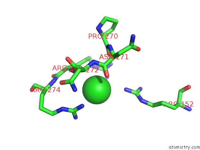
Mono view
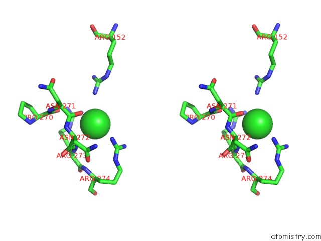
Stereo pair view
Mono view
Stereo pair view
A full contact list of Chlorine with other atoms in the Cl binding
site number 2 of Crystal Structure of Penicillin-Binding Protein 3 From Pseudomonas Aeruginosa in Complex with Piperacillin within 5.0Å range:
|
Reference:
S.S.Van Berkel,
J.E.Nettleship,
I.K.Leung,
J.Brem,
H.Choi,
D.I.Stuart,
T.D.Claridge,
M.A.Mcdonough,
R.J.Owens,
J.Ren,
C.J.Schofield.
Binding of (5S)-Penicilloic Acid to Penicillin Binding Protein 3. Acs Chem.Biol. V. 8 2112 2013.
ISSN: ISSN 1554-8929
PubMed: 23899657
DOI: 10.1021/CB400200H
Page generated: Fri Jul 11 17:53:22 2025
ISSN: ISSN 1554-8929
PubMed: 23899657
DOI: 10.1021/CB400200H
Last articles
Na in 1VI6Na in 1VKG
Na in 1VMJ
Na in 1VMH
Na in 1VMF
Na in 1VLM
Na in 1VK1
Na in 1VIZ
Na in 1VEL
Na in 1VE8