Chlorine »
PDB 4kwy-4lb8 »
4kxh »
Chlorine in PDB 4kxh: The X-Ray Crystal Structure of A Dimeric Variant of Human Pancreatic Ribonuclease
Enzymatic activity of The X-Ray Crystal Structure of A Dimeric Variant of Human Pancreatic Ribonuclease
All present enzymatic activity of The X-Ray Crystal Structure of A Dimeric Variant of Human Pancreatic Ribonuclease:
3.1.27.5;
3.1.27.5;
Protein crystallography data
The structure of The X-Ray Crystal Structure of A Dimeric Variant of Human Pancreatic Ribonuclease, PDB code: 4kxh
was solved by
A.Pica,
A.Merlino,
L.Mazzarella,
F.Sica,
with X-Ray Crystallography technique. A brief refinement statistics is given in the table below:
| Resolution Low / High (Å) | 19.82 / 2.70 |
| Space group | P 21 21 21 |
| Cell size a, b, c (Å), α, β, γ (°) | 71.506, 74.319, 128.533, 90.00, 90.00, 90.00 |
| R / Rfree (%) | 19.8 / 24.6 |
Other elements in 4kxh:
The structure of The X-Ray Crystal Structure of A Dimeric Variant of Human Pancreatic Ribonuclease also contains other interesting chemical elements:
| Sodium | (Na) | 3 atoms |
Chlorine Binding Sites:
The binding sites of Chlorine atom in the The X-Ray Crystal Structure of A Dimeric Variant of Human Pancreatic Ribonuclease
(pdb code 4kxh). This binding sites where shown within
5.0 Angstroms radius around Chlorine atom.
In total only one binding site of Chlorine was determined in the The X-Ray Crystal Structure of A Dimeric Variant of Human Pancreatic Ribonuclease, PDB code: 4kxh:
In total only one binding site of Chlorine was determined in the The X-Ray Crystal Structure of A Dimeric Variant of Human Pancreatic Ribonuclease, PDB code: 4kxh:
Chlorine binding site 1 out of 1 in 4kxh
Go back to
Chlorine binding site 1 out
of 1 in the The X-Ray Crystal Structure of A Dimeric Variant of Human Pancreatic Ribonuclease
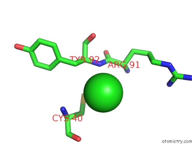
Mono view
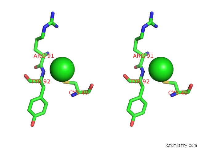
Stereo pair view
Mono view
Stereo pair view
A full contact list of Chlorine with other atoms in the Cl binding
site number 1 of The X-Ray Crystal Structure of A Dimeric Variant of Human Pancreatic Ribonuclease within 5.0Å range:
|
Reference:
A.Pica,
A.Merlino,
A.K.Buell,
T.P.Knowles,
E.Pizzo,
G.D'alessio,
F.Sica,
L.Mazzarella.
Three-Dimensional Domain Swapping and Supramolecular Protein Assembly: Insights From the X-Ray Structure of A Dimeric Swapped Variant of Human Pancreatic Rnase. Acta Crystallogr.,Sect.D V. 69 2116 2013.
ISSN: ISSN 0907-4449
PubMed: 24100329
DOI: 10.1107/S0907444913020507
Page generated: Fri Jul 11 18:04:41 2025
ISSN: ISSN 0907-4449
PubMed: 24100329
DOI: 10.1107/S0907444913020507
Last articles
Na in 4WOJNa in 4WOA
Na in 4WNW
Na in 4WO6
Na in 4WNV
Na in 4WMU
Na in 4WM5
Na in 4WM4
Na in 4WM3
Na in 4WM2