Chlorine »
PDB 4lr4-4lxd »
4ls6 »
Chlorine in PDB 4ls6: Crystal Structure of Beta-Ketoacyl-Acp Synthase II (Fabf) I108F Mutant From Bacillus Subtilis
Enzymatic activity of Crystal Structure of Beta-Ketoacyl-Acp Synthase II (Fabf) I108F Mutant From Bacillus Subtilis
All present enzymatic activity of Crystal Structure of Beta-Ketoacyl-Acp Synthase II (Fabf) I108F Mutant From Bacillus Subtilis:
2.3.1.179;
2.3.1.179;
Protein crystallography data
The structure of Crystal Structure of Beta-Ketoacyl-Acp Synthase II (Fabf) I108F Mutant From Bacillus Subtilis, PDB code: 4ls6
was solved by
F.Trajtenberg,
N.Larrieux,
A.Buschiazzo,
with X-Ray Crystallography technique. A brief refinement statistics is given in the table below:
| Resolution Low / High (Å) | 22.84 / 1.56 |
| Space group | P 21 21 21 |
| Cell size a, b, c (Å), α, β, γ (°) | 72.069, 87.974, 145.054, 90.00, 90.00, 90.00 |
| R / Rfree (%) | 14.9 / 17.2 |
Other elements in 4ls6:
The structure of Crystal Structure of Beta-Ketoacyl-Acp Synthase II (Fabf) I108F Mutant From Bacillus Subtilis also contains other interesting chemical elements:
| Potassium | (K) | 4 atoms |
Chlorine Binding Sites:
The binding sites of Chlorine atom in the Crystal Structure of Beta-Ketoacyl-Acp Synthase II (Fabf) I108F Mutant From Bacillus Subtilis
(pdb code 4ls6). This binding sites where shown within
5.0 Angstroms radius around Chlorine atom.
In total only one binding site of Chlorine was determined in the Crystal Structure of Beta-Ketoacyl-Acp Synthase II (Fabf) I108F Mutant From Bacillus Subtilis, PDB code: 4ls6:
In total only one binding site of Chlorine was determined in the Crystal Structure of Beta-Ketoacyl-Acp Synthase II (Fabf) I108F Mutant From Bacillus Subtilis, PDB code: 4ls6:
Chlorine binding site 1 out of 1 in 4ls6
Go back to
Chlorine binding site 1 out
of 1 in the Crystal Structure of Beta-Ketoacyl-Acp Synthase II (Fabf) I108F Mutant From Bacillus Subtilis
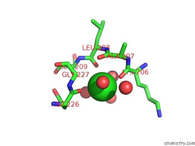
Mono view
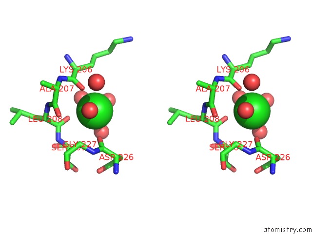
Stereo pair view
Mono view
Stereo pair view
A full contact list of Chlorine with other atoms in the Cl binding
site number 1 of Crystal Structure of Beta-Ketoacyl-Acp Synthase II (Fabf) I108F Mutant From Bacillus Subtilis within 5.0Å range:
|
Reference:
F.Trajtenberg,
S.Altabe,
N.Larrieux,
F.Ficarra,
D.De Mendoza,
A.Buschiazzo,
G.E.Schujman.
Structural Insights Into Bacterial Resistance to Cerulenin. Febs J. V. 281 2324 2014.
ISSN: ISSN 1742-464X
PubMed: 24641521
DOI: 10.1111/FEBS.12785
Page generated: Fri Jul 11 18:44:06 2025
ISSN: ISSN 1742-464X
PubMed: 24641521
DOI: 10.1111/FEBS.12785
Last articles
Mg in 2VSCMg in 2VPN
Mg in 2VRN
Mg in 2VPQ
Mg in 2VQD
Mg in 2VQ2
Mg in 2VPR
Mg in 2VPO
Mg in 2VP0
Mg in 2VOS