Chlorine »
PDB 4lxh-4m5g »
4m0g »
Chlorine in PDB 4m0g: The Crystal Structure of An Adenylosuccinate Synthetase From Bacillus Anthracis Str. Ames Ancestor.
Enzymatic activity of The Crystal Structure of An Adenylosuccinate Synthetase From Bacillus Anthracis Str. Ames Ancestor.
All present enzymatic activity of The Crystal Structure of An Adenylosuccinate Synthetase From Bacillus Anthracis Str. Ames Ancestor.:
6.3.4.4;
6.3.4.4;
Protein crystallography data
The structure of The Crystal Structure of An Adenylosuccinate Synthetase From Bacillus Anthracis Str. Ames Ancestor., PDB code: 4m0g
was solved by
K.Tan,
M.Zhou,
R.Zhang,
K.Kwon,
W.F.Anderson,
A.Joachimiak,
Midwest Centerfor Structural Genomics (Mcsg),
with X-Ray Crystallography technique. A brief refinement statistics is given in the table below:
| Resolution Low / High (Å) | 45.74 / 2.15 |
| Space group | P 41 21 2 |
| Cell size a, b, c (Å), α, β, γ (°) | 76.498, 76.498, 342.797, 90.00, 90.00, 90.00 |
| R / Rfree (%) | 19.2 / 24.3 |
Chlorine Binding Sites:
The binding sites of Chlorine atom in the The Crystal Structure of An Adenylosuccinate Synthetase From Bacillus Anthracis Str. Ames Ancestor.
(pdb code 4m0g). This binding sites where shown within
5.0 Angstroms radius around Chlorine atom.
In total only one binding site of Chlorine was determined in the The Crystal Structure of An Adenylosuccinate Synthetase From Bacillus Anthracis Str. Ames Ancestor., PDB code: 4m0g:
In total only one binding site of Chlorine was determined in the The Crystal Structure of An Adenylosuccinate Synthetase From Bacillus Anthracis Str. Ames Ancestor., PDB code: 4m0g:
Chlorine binding site 1 out of 1 in 4m0g
Go back to
Chlorine binding site 1 out
of 1 in the The Crystal Structure of An Adenylosuccinate Synthetase From Bacillus Anthracis Str. Ames Ancestor.
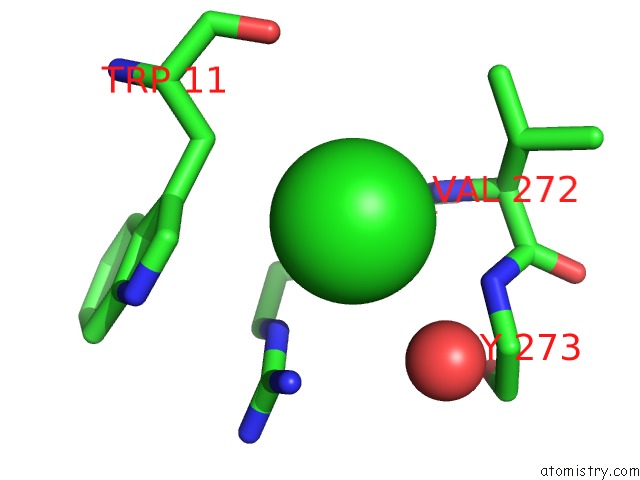
Mono view
Stereo pair view
Mono view
Stereo pair view
A full contact list of Chlorine with other atoms in the Cl binding
site number 1 of The Crystal Structure of An Adenylosuccinate Synthetase From Bacillus Anthracis Str. Ames Ancestor. within 5.0Å range:
|
Reference:
K.Tan,
M.Zhou,
R.Zhang,
K.Kwon,
W.F.Anderson,
A.Joachimiak.
The Crystal Structure of An Adenylosuccinate Synthetase From Bacillus Anthracis Str. Ames Ancestor. To Be Published.
Page generated: Fri Jul 11 18:52:03 2025
Last articles
Ni in 1EM0Ni in 1ELW
Ni in 1EJX
Ni in 1ELR
Ni in 1EK0
Ni in 1EJW
Ni in 1EJV
Ni in 1EJU
Ni in 1EJT
Ni in 1EJS