Chlorine »
PDB 4lxh-4m5g »
4m3k »
Chlorine in PDB 4m3k: Structure of A Single Domain Camelid Antibody Fragment Cab-H7S in Complex with the Blap Beta-Lactamase From Bacillus Licheniformis
Enzymatic activity of Structure of A Single Domain Camelid Antibody Fragment Cab-H7S in Complex with the Blap Beta-Lactamase From Bacillus Licheniformis
All present enzymatic activity of Structure of A Single Domain Camelid Antibody Fragment Cab-H7S in Complex with the Blap Beta-Lactamase From Bacillus Licheniformis:
3.5.2.6;
3.5.2.6;
Protein crystallography data
The structure of Structure of A Single Domain Camelid Antibody Fragment Cab-H7S in Complex with the Blap Beta-Lactamase From Bacillus Licheniformis, PDB code: 4m3k
was solved by
C.Pain,
F.Kerff,
R.Herman,
E.Sauvage,
S.Preumont,
P.Charlier,
M.Dumoulin,
with X-Ray Crystallography technique. A brief refinement statistics is given in the table below:
| Resolution Low / High (Å) | 36.90 / 1.48 |
| Space group | P 1 21 1 |
| Cell size a, b, c (Å), α, β, γ (°) | 51.818, 42.940, 74.818, 90.00, 105.29, 90.00 |
| R / Rfree (%) | 16.1 / 19.5 |
Chlorine Binding Sites:
The binding sites of Chlorine atom in the Structure of A Single Domain Camelid Antibody Fragment Cab-H7S in Complex with the Blap Beta-Lactamase From Bacillus Licheniformis
(pdb code 4m3k). This binding sites where shown within
5.0 Angstroms radius around Chlorine atom.
In total 2 binding sites of Chlorine where determined in the Structure of A Single Domain Camelid Antibody Fragment Cab-H7S in Complex with the Blap Beta-Lactamase From Bacillus Licheniformis, PDB code: 4m3k:
Jump to Chlorine binding site number: 1; 2;
In total 2 binding sites of Chlorine where determined in the Structure of A Single Domain Camelid Antibody Fragment Cab-H7S in Complex with the Blap Beta-Lactamase From Bacillus Licheniformis, PDB code: 4m3k:
Jump to Chlorine binding site number: 1; 2;
Chlorine binding site 1 out of 2 in 4m3k
Go back to
Chlorine binding site 1 out
of 2 in the Structure of A Single Domain Camelid Antibody Fragment Cab-H7S in Complex with the Blap Beta-Lactamase From Bacillus Licheniformis
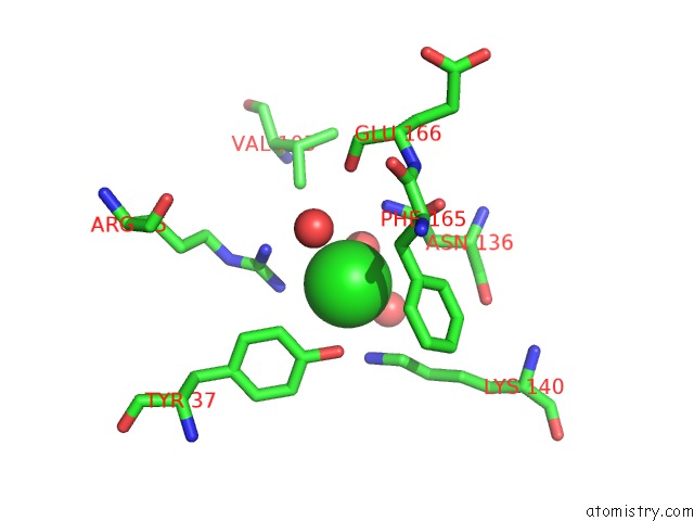
Mono view
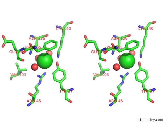
Stereo pair view
Mono view
Stereo pair view
A full contact list of Chlorine with other atoms in the Cl binding
site number 1 of Structure of A Single Domain Camelid Antibody Fragment Cab-H7S in Complex with the Blap Beta-Lactamase From Bacillus Licheniformis within 5.0Å range:
|
Chlorine binding site 2 out of 2 in 4m3k
Go back to
Chlorine binding site 2 out
of 2 in the Structure of A Single Domain Camelid Antibody Fragment Cab-H7S in Complex with the Blap Beta-Lactamase From Bacillus Licheniformis
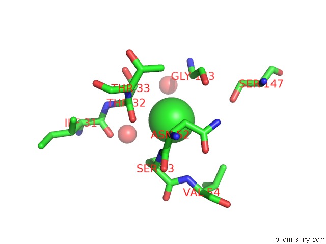
Mono view
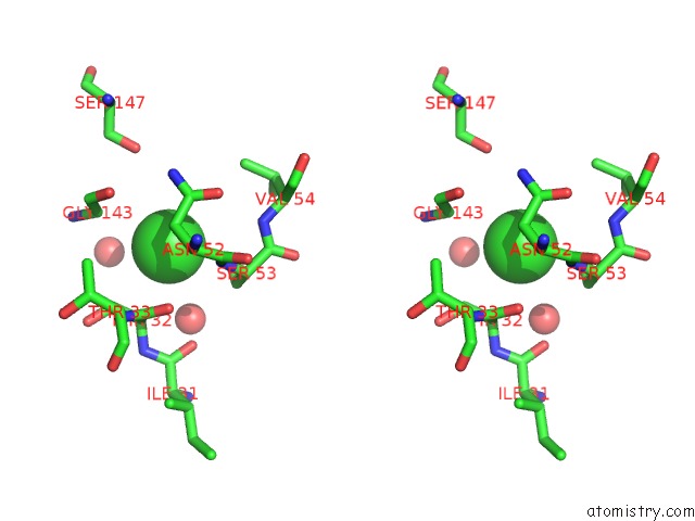
Stereo pair view
Mono view
Stereo pair view
A full contact list of Chlorine with other atoms in the Cl binding
site number 2 of Structure of A Single Domain Camelid Antibody Fragment Cab-H7S in Complex with the Blap Beta-Lactamase From Bacillus Licheniformis within 5.0Å range:
|
Reference:
C.Pain,
A.Cosolo,
S.Preumont,
N.Scarafone,
D.Thorn,
R.Herman,
H.Spiegel,
E.Pardon,
A.Matagne,
P.Charlier,
J.Steyaert,
C.Damblon,
F.Kerff,
G.Esposito,
M.Dumoulin.
Probing the Mechanism of Aggregation of Polyq Model Proteins with Camelid Heavy-Chain Antibody Fragments To Be Published.
Page generated: Fri Jul 11 18:55:12 2025
Last articles
Na in 9OW2Na in 9OS1
Na in 9ORZ
Na in 9OS0
Na in 9NH3
Na in 9ORW
Na in 9ORV
Na in 9NGR
Na in 9NGQ
Na in 9NGS