Chlorine »
PDB 4m5h-4mdq »
4mdq »
Chlorine in PDB 4mdq: Structure of A Novel Submicromolar MDM2 Inhibitor
Protein crystallography data
The structure of Structure of A Novel Submicromolar MDM2 Inhibitor, PDB code: 4mdq
was solved by
M.Bista,
G.Popowicz,
T.A.Holak,
with X-Ray Crystallography technique. A brief refinement statistics is given in the table below:
| Resolution Low / High (Å) | 46.35 / 2.12 |
| Space group | P 65 2 2 |
| Cell size a, b, c (Å), α, β, γ (°) | 53.520, 53.520, 122.270, 90.00, 90.00, 120.00 |
| R / Rfree (%) | 18.5 / 22.1 |
Chlorine Binding Sites:
The binding sites of Chlorine atom in the Structure of A Novel Submicromolar MDM2 Inhibitor
(pdb code 4mdq). This binding sites where shown within
5.0 Angstroms radius around Chlorine atom.
In total 2 binding sites of Chlorine where determined in the Structure of A Novel Submicromolar MDM2 Inhibitor, PDB code: 4mdq:
Jump to Chlorine binding site number: 1; 2;
In total 2 binding sites of Chlorine where determined in the Structure of A Novel Submicromolar MDM2 Inhibitor, PDB code: 4mdq:
Jump to Chlorine binding site number: 1; 2;
Chlorine binding site 1 out of 2 in 4mdq
Go back to
Chlorine binding site 1 out
of 2 in the Structure of A Novel Submicromolar MDM2 Inhibitor
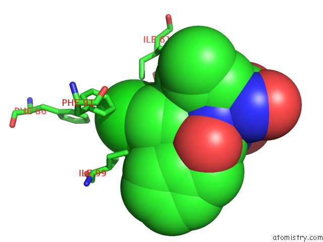
Mono view
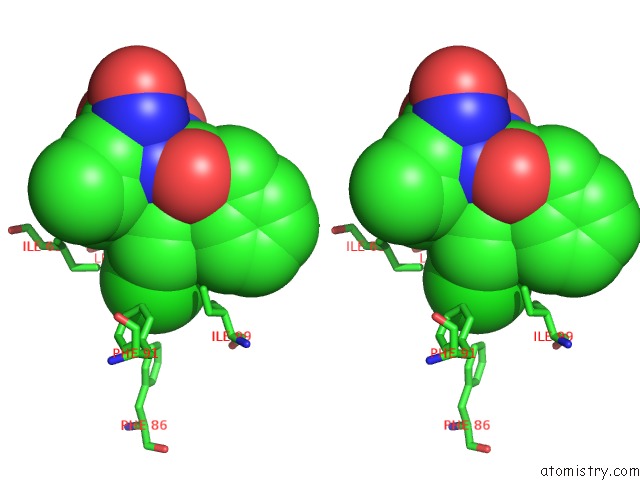
Stereo pair view
Mono view
Stereo pair view
A full contact list of Chlorine with other atoms in the Cl binding
site number 1 of Structure of A Novel Submicromolar MDM2 Inhibitor within 5.0Å range:
|
Chlorine binding site 2 out of 2 in 4mdq
Go back to
Chlorine binding site 2 out
of 2 in the Structure of A Novel Submicromolar MDM2 Inhibitor
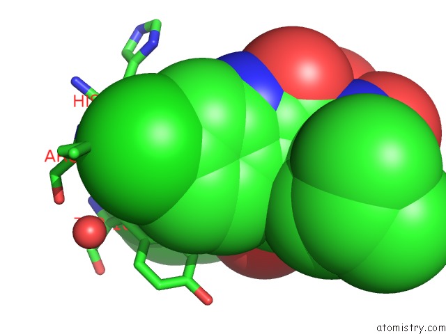
Mono view
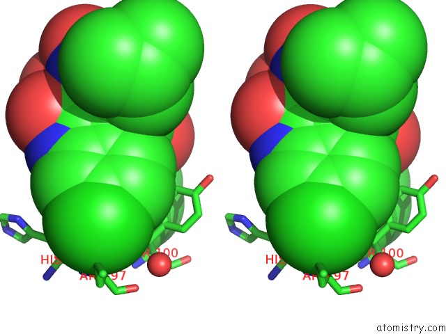
Stereo pair view
Mono view
Stereo pair view
A full contact list of Chlorine with other atoms in the Cl binding
site number 2 of Structure of A Novel Submicromolar MDM2 Inhibitor within 5.0Å range:
|
Reference:
M.Bista,
S.Wolf,
K.Khoury,
K.Kowalska,
Y.Huang,
E.Wrona,
M.Arciniega,
G.M.Popowicz,
T.A.Holak,
A.Domling.
Transient Protein States in Designing Inhibitors of the MDM2-P53 Interaction. Structure V. 21 2143 2013.
ISSN: ISSN 0969-2126
PubMed: 24207125
DOI: 10.1016/J.STR.2013.09.006
Page generated: Fri Jul 11 19:02:17 2025
ISSN: ISSN 0969-2126
PubMed: 24207125
DOI: 10.1016/J.STR.2013.09.006
Last articles
Mg in 6B5EMg in 6B4O
Mg in 6AZ1
Mg in 6B4J
Mg in 6B4K
Mg in 6B3E
Mg in 6B3V
Mg in 6B3K
Mg in 6B1N
Mg in 6B1M