Chlorine »
PDB 4mlb-4ms4 »
4mqf »
Chlorine in PDB 4mqf: Crystal Structure of the Extracellular Domain of Human Gaba(B) Receptor Bound to the Antagonist 2-Hydroxysaclofen
Protein crystallography data
The structure of Crystal Structure of the Extracellular Domain of Human Gaba(B) Receptor Bound to the Antagonist 2-Hydroxysaclofen, PDB code: 4mqf
was solved by
Y.Geng,
M.Bush,
L.Mosyak,
F.Wang,
Q.R.Fan,
with X-Ray Crystallography technique. A brief refinement statistics is given in the table below:
| Resolution Low / High (Å) | 32.50 / 2.22 |
| Space group | P 1 21 1 |
| Cell size a, b, c (Å), α, β, γ (°) | 70.280, 112.630, 73.220, 90.00, 97.79, 90.00 |
| R / Rfree (%) | 20.7 / 23.4 |
Chlorine Binding Sites:
The binding sites of Chlorine atom in the Crystal Structure of the Extracellular Domain of Human Gaba(B) Receptor Bound to the Antagonist 2-Hydroxysaclofen
(pdb code 4mqf). This binding sites where shown within
5.0 Angstroms radius around Chlorine atom.
In total only one binding site of Chlorine was determined in the Crystal Structure of the Extracellular Domain of Human Gaba(B) Receptor Bound to the Antagonist 2-Hydroxysaclofen, PDB code: 4mqf:
In total only one binding site of Chlorine was determined in the Crystal Structure of the Extracellular Domain of Human Gaba(B) Receptor Bound to the Antagonist 2-Hydroxysaclofen, PDB code: 4mqf:
Chlorine binding site 1 out of 1 in 4mqf
Go back to
Chlorine binding site 1 out
of 1 in the Crystal Structure of the Extracellular Domain of Human Gaba(B) Receptor Bound to the Antagonist 2-Hydroxysaclofen
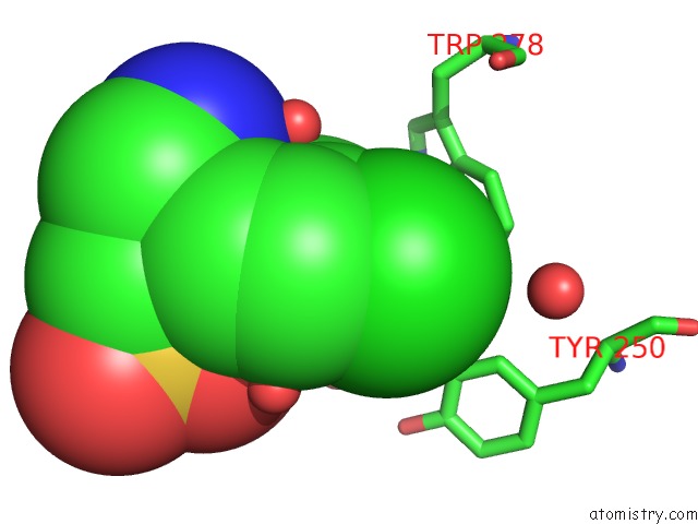
Mono view
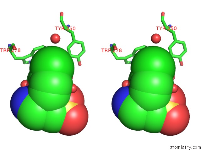
Stereo pair view
Mono view
Stereo pair view
A full contact list of Chlorine with other atoms in the Cl binding
site number 1 of Crystal Structure of the Extracellular Domain of Human Gaba(B) Receptor Bound to the Antagonist 2-Hydroxysaclofen within 5.0Å range:
|
Reference:
Y.Geng,
M.Bush,
L.Mosyak,
F.Wang,
Q.R.Fan.
Structural Mechanism of Ligand Activation in Human Gaba(B) Receptor. Nature V. 504 254 2013.
ISSN: ISSN 0028-0836
PubMed: 24305054
DOI: 10.1038/NATURE12725
Page generated: Fri Jul 11 19:16:05 2025
ISSN: ISSN 0028-0836
PubMed: 24305054
DOI: 10.1038/NATURE12725
Last articles
I in 4PX3I in 4PX2
I in 4P9T
I in 4PNX
I in 4PVQ
I in 4PVG
I in 4PGC
I in 4PNS
I in 4P4Z
I in 4P1E