Chlorine »
PDB 4msh-4myq »
4mvt »
Chlorine in PDB 4mvt: Crystal Structure of Sumo E3 Ligase PIAS3
Protein crystallography data
The structure of Crystal Structure of Sumo E3 Ligase PIAS3, PDB code: 4mvt
was solved by
A.Dong,
J.Hu,
Y.Li,
W.Tempel,
C.Bountra,
C.H.Arrowsmith,
A.M.Edwards,
Y.Tong,
Structural Genomics Consortium (Sgc),
with X-Ray Crystallography technique. A brief refinement statistics is given in the table below:
| Resolution Low / High (Å) | 48.04 / 2.30 |
| Space group | P 1 |
| Cell size a, b, c (Å), α, β, γ (°) | 55.450, 85.440, 89.530, 83.08, 86.57, 86.14 |
| R / Rfree (%) | 23.8 / 27 |
Other elements in 4mvt:
The structure of Crystal Structure of Sumo E3 Ligase PIAS3 also contains other interesting chemical elements:
| Zinc | (Zn) | 4 atoms |
Chlorine Binding Sites:
The binding sites of Chlorine atom in the Crystal Structure of Sumo E3 Ligase PIAS3
(pdb code 4mvt). This binding sites where shown within
5.0 Angstroms radius around Chlorine atom.
In total 2 binding sites of Chlorine where determined in the Crystal Structure of Sumo E3 Ligase PIAS3, PDB code: 4mvt:
Jump to Chlorine binding site number: 1; 2;
In total 2 binding sites of Chlorine where determined in the Crystal Structure of Sumo E3 Ligase PIAS3, PDB code: 4mvt:
Jump to Chlorine binding site number: 1; 2;
Chlorine binding site 1 out of 2 in 4mvt
Go back to
Chlorine binding site 1 out
of 2 in the Crystal Structure of Sumo E3 Ligase PIAS3
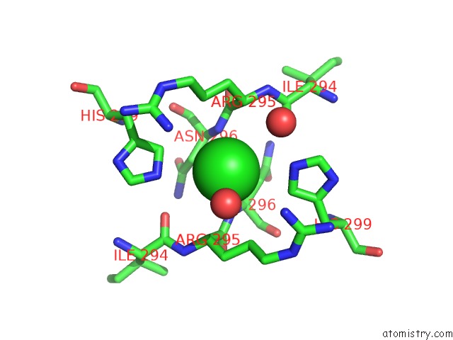
Mono view
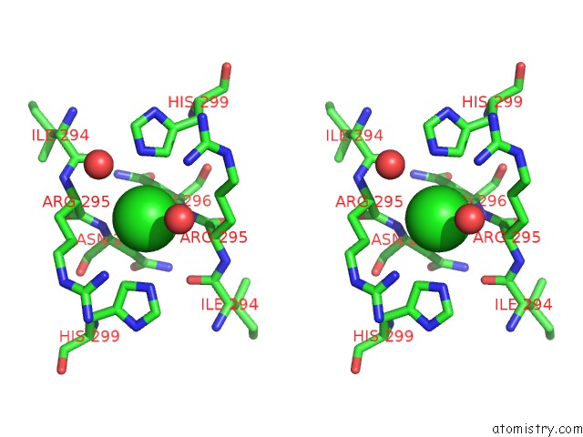
Stereo pair view
Mono view
Stereo pair view
A full contact list of Chlorine with other atoms in the Cl binding
site number 1 of Crystal Structure of Sumo E3 Ligase PIAS3 within 5.0Å range:
|
Chlorine binding site 2 out of 2 in 4mvt
Go back to
Chlorine binding site 2 out
of 2 in the Crystal Structure of Sumo E3 Ligase PIAS3
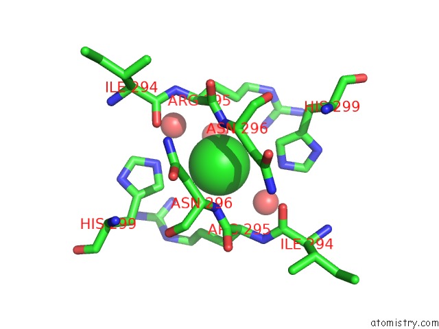
Mono view
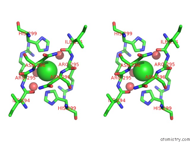
Stereo pair view
Mono view
Stereo pair view
A full contact list of Chlorine with other atoms in the Cl binding
site number 2 of Crystal Structure of Sumo E3 Ligase PIAS3 within 5.0Å range:
|
Reference:
J.Hu,
A.Dong,
Y.Li,
W.Tempel,
C.Bountra,
C.H.Arrowsmith,
A.M.Edwards,
Y.Tong,
Structural Genomics Consortium (Sgc).
Crystal Structure of Sumo E3 Ligase PIAS3 To Be Published.
Page generated: Fri Jul 11 19:18:58 2025
Last articles
Mg in 9JNTMg in 9JNP
Mg in 9JN3
Mg in 9JMA
Mg in 9JKP
Mg in 9JJW
Mg in 9JJV
Mg in 9JI2
Mg in 9JFJ
Mg in 9J89