Chlorine »
PDB 4n9a-4ngr »
4n9m »
Chlorine in PDB 4n9m: Joint Neutron/X-Ray Structure of Urate Oxidase in Complex with 8- Hydroxyxanthine
Enzymatic activity of Joint Neutron/X-Ray Structure of Urate Oxidase in Complex with 8- Hydroxyxanthine
All present enzymatic activity of Joint Neutron/X-Ray Structure of Urate Oxidase in Complex with 8- Hydroxyxanthine:
1.7.3.3;
1.7.3.3;
Protein crystallography data
The structure of Joint Neutron/X-Ray Structure of Urate Oxidase in Complex with 8- Hydroxyxanthine, PDB code: 4n9m
was solved by
E.Oksanen,
M.P.Blakeley,
M.Budayova-Spano,
with X-Ray Crystallography technique. A brief refinement statistics is given in the table below:
| Resolution Low / High (Å) | N/A / 2.30 |
| Space group | I 2 2 2 |
| Cell size a, b, c (Å), α, β, γ (°) | 80.180, 96.260, 105.510, 90.00, 90.00, 90.00 |
| R / Rfree (%) | 16.7 / 18.9 |
Other elements in 4n9m:
The structure of Joint Neutron/X-Ray Structure of Urate Oxidase in Complex with 8- Hydroxyxanthine also contains other interesting chemical elements:
| Sodium | (Na) | 1 atom |
Chlorine Binding Sites:
The binding sites of Chlorine atom in the Joint Neutron/X-Ray Structure of Urate Oxidase in Complex with 8- Hydroxyxanthine
(pdb code 4n9m). This binding sites where shown within
5.0 Angstroms radius around Chlorine atom.
In total only one binding site of Chlorine was determined in the Joint Neutron/X-Ray Structure of Urate Oxidase in Complex with 8- Hydroxyxanthine, PDB code: 4n9m:
In total only one binding site of Chlorine was determined in the Joint Neutron/X-Ray Structure of Urate Oxidase in Complex with 8- Hydroxyxanthine, PDB code: 4n9m:
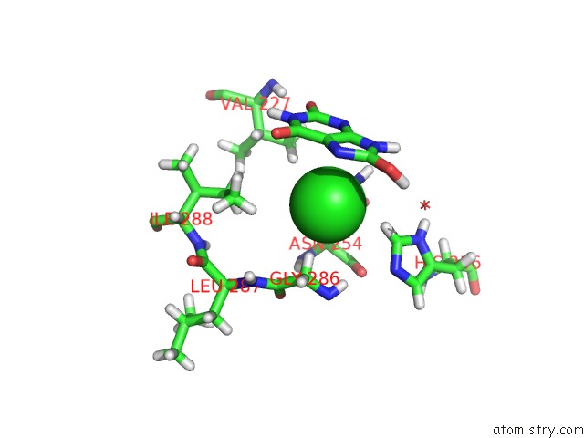
Chlorine binding site 1 out of 1 in 4n9m
Go back to
Chlorine binding site 1 out
of 1 in the Joint Neutron/X-Ray Structure of Urate Oxidase in Complex with 8- Hydroxyxanthine
Mono view

Stereo pair view
Mono view
Stereo pair view
A full contact list of Chlorine with other atoms in the Cl binding
site number 1 of Joint Neutron/X-Ray Structure of Urate Oxidase in Complex with 8- Hydroxyxanthine within 5.0Å range:
|
Reference:
E.Oksanen,
M.P.Blakeley,
M.El-Hajji,
U.Ryde,
M.Budayova-Spano.
The Neutron Structure of Urate Oxidase Resolves A Long-Standing Mechanistic Conundrum and Reveals Unexpected Changes in Protonation. Plos One V. 9 86651 2014.
ISSN: ESSN 1932-6203
PubMed: 24466188
DOI: 10.1371/JOURNAL.PONE.0086651
Page generated: Fri Jul 11 19:31:45 2025
ISSN: ESSN 1932-6203
PubMed: 24466188
DOI: 10.1371/JOURNAL.PONE.0086651
Last articles
K in 9GN7K in 9GN6
K in 9GOY
K in 9GKZ
K in 9GLB
K in 9GN1
K in 9GKY
K in 9GL0
K in 9GL1
K in 9GKW